Images in Oncology via PET/CT
Your oncology lecture has numerous links to this page giving you examples of what different types o carcinomas look like when imaged with PET/CT, usually FDG is on-board.
- Pancoast Tumor
- This is an example of NSCLC tumor that has actually developed into a pancoast tumor (red arrow). This name is given by the structural design of the tumor and has nothing to do with its physiology
- Pink arrows show regional lymph node involvement
- Given the fact that there is lymph node involvement would this person be a candidate for surgery? Answer
- This image was taken from a series of images that can be reviewed at: http://rad.usuhs.edu/medpix/radpix.html?mode=single&recnum=4588&table=&srchstr=&search= (this may not be active)
- Second example of NSCLC tumor
- Primary tumor is defined as A, B identifies mediastinal lymph node involvement
- Dotted lines show on the whole body that cancer is involvement in the cervical and supraclavicular lymph nodes
- Adenocarcinoma
- An example of a primary adenocarcinoma of the lung
- This is an excellent example of a candidate for surgery since the only area that FDG avid is in the tumor. Had there been lymph node involvement that surgery would not have been the best treatment option because the cancer had gone beyond the tumor bed. Radiation therapy and/or chemotherapy would have be the next course of treatment
- Bronchoalveolar
- This is a 62 year old female with significant involvement of bronchoalveolar carcinoma
- While arrow defines the primary the black arrow point two of the many metastatic lesions seen in the bone tissue
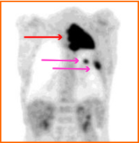
- Squamous Cell Lung Cancer
- This cancerous squamous cell invades upper pole of the lung
- Metastatic lymph node involvement can also be identified
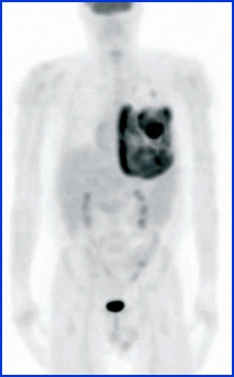
- Mesotheloioma
- This is an example of pleural mesothelioma
- Usually the cause of this cancer is related to the excessive exposure to asbestos
- Breast
 1
1
- Papillary breast cancer - Patient underwent left quadrantectomy followed by radiation therapy. Two years later a PET scan revealed the above disease
- Diffused gastric uptake then apparently is not caner related
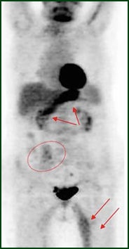
- Infection and/or trauma in her left thigh
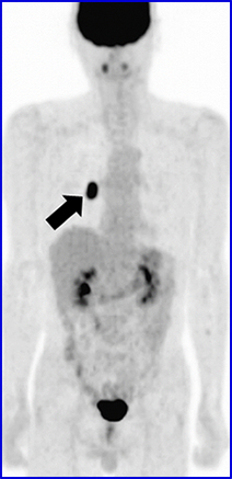
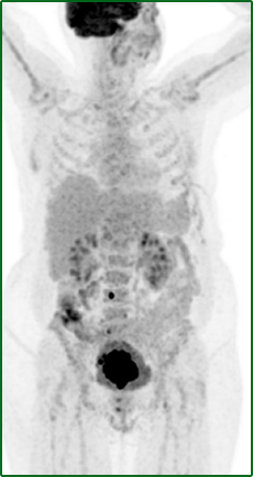
- Circle is is the right iliac which is a metastatic lesion
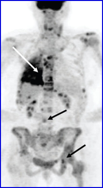
- Thyroid cancer
- Metastatic involvement is noted in the neck and mediastinum specifically within the lymphatic system
- http://www.csmc.edu/6982.html (3/15 link was off line)

http://www.medscape.com/viewarticle/586604_3
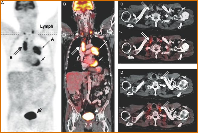
- Metastatic Lymph nodes
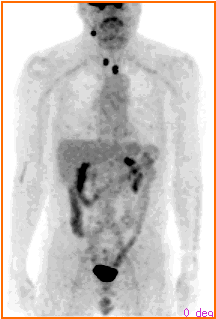
- 72-year-old female of endometrial adenocarcinoma. Demonstrates increased uptake in the entire uterus, consistent with hypermetabolic malignancy (arrow heads). In addition, there are multiple FDG avid lymph nodes along the pelvic wall bilaterally (arrows), left paraaortic nodal basin (arrow A), right inguinal lymph node (arrow B). The findings are consistent with lymph node metastasis. (quoted from article)
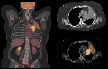
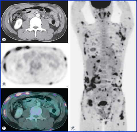
- Liver and Pancreatic Cancers
- FDG identifies cholagiocarcinoma with no metastatic involvement
- Pancreatic cancer is initially identified with PET/CT FDG. With the application of contrast fused volume-CT rendering the superior mesenteric artery structure with FDG uptake identifying tumor at the pancreatic head
- GU Cancers
- Bladder cancer - There is some limitation to this exam since FDG is excreted by the kidneys. For this reason, when imaging the bladder: force hydration and diuretics, and a Foley catheter might help improve imaging in this area
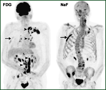
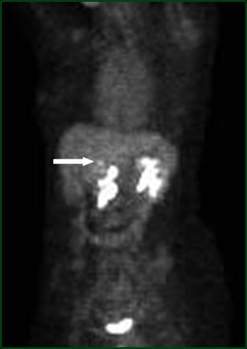
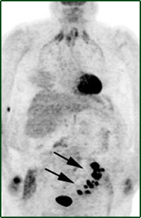
- Prostate cancer - FDG and 18NaF images were taken on the same patient. Remember Na18F shows bone turnover whereas FDG goes to metabolic uptake. While bone lesions are seen on both, each exam also shows its limitations of finding disease
- FDG shows additional lymphatic involvement
- Na18F identifies additional lesions in the bone
http://journal.frontiersin.org/article/10.3389/fonc.2013.00034/full
- Cervical cancer may appear to be only at its primary, however, there may also be some minor bone involvement
- Lymphoma
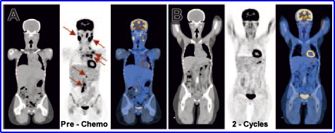
- Subcutaneous T-cell
- This scan was completed with FDG for initial staging of disease
http://www.intechopen.com/books/selected-topics-on-computed-tomography/molecular-imaging
- While follow-up images are not available, after three cycles of chemotherapy there was complete remission of disease
Return to PET Oncology Lecture
1. Whole Body FDG PET Imaging in Oncology by Rambaldi, pp 217-218, Springer