Spleen - Anatomy, Physiology, and Imaging
- Anatomy
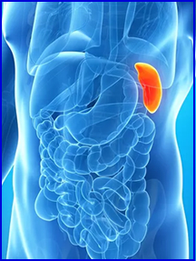
- The spleen is an oval shaped mass of lymphatic tissue located in the left upper quadrant (LUQ)
- It is situated between the fundus of the stomach and the left half of the diaphragm
- It is lateral and superior to the left kidney
- The adult spleen measures approximately 14 cm in length and weighs about 200 grams
- The spleen is hematopoietic system that includes the RES
- The best images of the spleen are:
- Left lateral
- Posterior
- Physiology
- The spleen has the capacity to increase blood flow through portal circulation and/or by undergo reticulated cell hyperplasia
- The spleen is a “reservoir” of blood elements
- The organ produces/store antibodies and platelets
- Components of blood
- RBC - Erythrocytes
- Hematocrit is the percent volume of RBC in the vascular pool, normal range is between 40 - 45%
- Production is controlled by erythropoietin, which is a hormone produced mostly by the kidneys
- An essential component of hemoglobin is the regulation of oxygen and carbon dioxide
- Average life expectancy of RBCs is 120 days
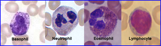
- WBC - Leukocytes
- On the average there is 1% circulating in the vascular pool.
- There are four major subgroups
- Neutrophils are the most plentiful (55 - 70%)
- Defense against bacterial and fungal injections
- Phagocytize bacteria
- Seen first when there is an acute infection
- Usually lives for about a day
- Eosinophil granulocytes
- Attacks parasitic infections
- Respond to allergic type reaction
- Allergies - asthma, hay fever, etc.
- Basophil
- Response to antigens and allergic reactions
- Releases histamine, which in turn causes vasodilation to increased blood flow to the area in need. It can, in extreme cases cause anaphylaxis response
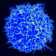
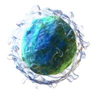
- Lymphocytes has to sub groups
T Lymphocyte
|
B Lymphocyte
|
- T lymphocytes - is what attacks antigens in the body and also has some regulatory functions with other immune cells
- B lymphocytes - makes the antibodies that attack the invading antigen
- Platelets - thrombocytes
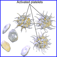
- Are fragments of cells which are used in the clotting process (coagulation) when injury occurs and/or if blood is leaking outside the vascular pool. It forms a fibrin clot allowing a healing process to occur
- The "10-Step Process of Blood Coagulation| Thrombocytes"
- The spleen can be a site of sequestration for any type of blood element
- Hypersplenism results in early and excessive trapping of blood cell elements. The spleen is a site of red cell elimination and destruction. If there is an excessive amount of trapping, splenectomy is sometimes recommended or the administration of Na32PO4-. Following splenectomy, the liver will take over the normal removal of blood cell elements. In nuclear medicine, an invitro test "Red Cell Survival" can be used to assess excessive elimination of RBCs
- The spleen removes red blood cells from circulation by culling and pitting
- Culling is a process where red cells (or their components) are removed from circulation when they become damaged or worn-out
- RBCs do not have nuclei or related DNA components, but there are certain diseases that cause nuclei or its remnants to be produced within the red cells. Culling removes this DNA by removing the RBC
- Furthermore, misshapen RBCs can be created. Hence the spleen's response is to destroy these cells in the same process
- Culling occurs as blood is filtered by the spleen through multiple layers of vessels (arterial and venous) with the assistance of macrophages
- Pitting is another process that occurs within the spleen, however, instead of removing RBCs, it repairs them. This can occur by the removal of certain DNA remnants or inclusion within or on the cell, As an example malaria causes inclusions within the RBC and pitting may remove some of those inclusions. This allows the body to put RBCs back into circulation
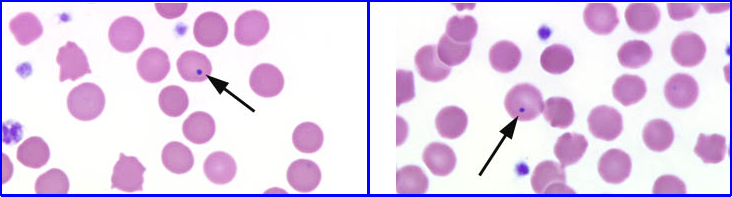
- Howell-Jolly Bodies are seen as inclusions and/or considered nuclei remnants. The spleen will either cull or pit these components. This is usually seen in patients with severe hemolytic anemia, dysfunction spleen, or has splenomegaly
- When removing particles think of the size - (<1μ) such as sulfur colloid (remnants) being removed by the spleen, however, larger particles, such as, labeled and denatured red blood cells (about 7μ) can be removed or sequestered. The process of removal would be known as __________?
- Another role the spleen has is that lymphocyte production of antibodies (occurring within the spleen) in response to foreign antigens. Note - In the cases where no spleen is present (or not functioning) the patient has at increased risk of infection.
Pathophysiology and disease
- Splenomegaly
- Disease causes enlargement of the organ
- Because it is an RES organ, acute and chronic infectious diseases as well as non- infectious inflammatory diseases cause splenomegaly
- Associated diseases usually include tuberculosis, infectious mononucleosis, subacute bacterial endocarditis, and septicemia. Leukemia may cause obstruction or excess cell formation which will result in splenomegaly. Benign and malignant infiltrate diseases cause splenomegaly and defects associated with RBC
- Spleen trauma
- Blunt trauma to the abdomen or to the lower rib cage may cause contusion or laceration to the spleen
- May result in excessive internal bleeding
- Even after the spleen has been removed, splenetic remnants have been found that was caused by trauma. Visualization of spleen remnants can be noted in colloid images in areas not associated with spleen or liver uptake
- Hyposplenism
- Surgical removal or splenetic infarctions cause hyposplenism, which may be responsible for future serious illness
- Causes increase in blood elements
- Platelet counts may elevate since the spleen is a platelet reservoir
Spleen imaging via nuclear medicine
- Indications for imaging
- Depiction of size, shape and position of the spleen
- Differentiation of LUQ masses
- Visualization of infiltration by such diseases as Hodgkin's’s (splenic lymphoma), reticulum cell sarcoma, or metastatic melanoma
- Evaluation of the spleen for possible damage resulting from trauma
- Accessory spleen or regeneration
Radiopharmaceutical
- The use of 99mTc sulfur colloid can be used and has been discussed in a previous lecture. The dose is 5 mCi
- Another approach is the use of 99mTc labeled RBC which requires denaturing of the RBCs
- Procedure for labeling and denaturing RBCs
- First label the RBCs - suggested method: use UltraTag (Ultratag RBC FDA Package Insert.pdf)
- Ultratag package insert
- Once the RBCs have been labeled add saline to 99mTc-RBC vial
- Place into water bath at 49o to 50o C
- Heat for 20 minutes
- Remove and let cool prior to administration
- The dose is now ready for IV administration - dose 10 - 15 mCi
- Start imaging 30 to 120 minutes post dose
- Take ANT, POST, LPO, and LAO imaging
- 750k counts
- SPECT may also be suggested
- If accessory spleen is expected then image the entire abdominal area
- If spleen trauma with diaphragmatic tearing is a concern then image the thoracic cavity
- The spleen will remove damaged red cells because denaturing changes the shape from biconcave disks to a spherical type object. In addition the cell membrane develops knobby projection
Imaging Protocol
- Collimation - LE Gap or LEHR
- Matrix size - 256 x 256
- Counts/image - 800 to 1000k (consider camera diameter)
- Images
- Anterior
- Anterior w/ marker,
- L-Lateral
- LAO
- LPO
- Posterior
- SPECT
- Matrix - 64 or 128
- Degrees - 360
- Type of orbit - elliptical or body contour
- 60 or 120 steps
- Maximum acquisition - 30 minute
Diseases
- Benign tumor known as a congenital hemangioma may be seen as space occupying lesions. Sometimes polycystic disease may involve the spleen
- Trauma typically caused by MVA will show intrasplenic hematoma that appear as a space occupying lesion. Subcapsular hematoma may show a deformed splenetic image. This may lead or cause ...
- Capsular rupture where internal bleeding from the trauma. Splenetic tissue may actually be displaced to another area of the body. Abnormal contour or a small section will be missing from the spleen form the spleen may be visualized
- Trauma may also cause accessory spleen
- Hematologic disease caused by acute and chronic leukemia will show splenetic enlargement
- Hodgkin's’s and Non-Hodgkin’s disease may show space occupying lesions. Spleen may or may not be enlarged
- Hemolytic anemia is indicated by spleen enlargement
- Liver disease causes other areas of the RES to take over that results in enlargement of the spleen with increased radiopharmaceutical shift to the spleen and bone marrow, colloidal shift
- Metastatic disease of the spleen is unusual, however, if seen there usually will be small space occupying lesions
- Sickle cell causes space occupying lesions which are due to micro infarcts
- Systemic infectious diseases will result in splenetic enlargement
Case presentations
-
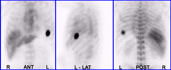
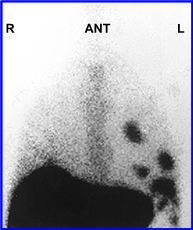
This patient was referred to the nuclear medicine department following an MVA that occurred two weeks prior to this study. The patient was injected with 5 mCi of 99mTc-sulfur colloid and imaged. The remarkable aspect of this scan is the increased amount of active noted in the area of the right pleural cavity. This was caused by severe trauma to the abdomen resulting in a small portion of the spleen to tear away, passing through an opening in the diaphragm. This accessory spleen has embedded itself along the muscular wall of the pleural cavity and establish flow blood as indicated in the images above. Technically this is known as an accessory spleen.
http://www.rah.sa.gov.au/nucmed/nm_linktous.htm
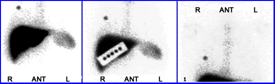
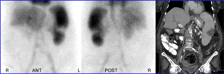
99mTc Labeled RBCs denatured by heating were injected into a patient for evaluation of spleen regeneration (splenectomy was completed on patient at some unknown date in the past). Twenty minutes post injection splenetic tissue is noted in the LUQ, identifying regenerated tissue. This is sometimes referred to as splenunculi which means accessory spleen. The following article discusses splenunculi in more detail medind.nic.in/jae/t02/i1/jaet02i1p70.pdf (this link is no longer active)
-
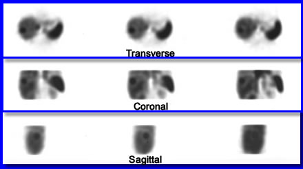
Not directly related to spleen imaging is another aspect of applying labeled RBCs (not denatured). Cavernous Hemangioma is a benign tumor that usually resides in the right lobe of the liver. As noted above, these SPECT images show an increased area of red cell accumulation. Cavernous Hemangioma "craves" red cells and as a result, over time will pick up enough labeled RBCs to be visualized.
A 76 year old male complained of continuous fever and had a splenectomy during WWII because of a gunshot wound to the abdomen. This case is presented at Medscape.
A 76 year old female with a history of multiple complications which included polycythemia Vera along with melena and hematemesis. After the injection of denatured RBCs a splenic infarct was discovered. CT scan further confirmed this diagnosis.
Return to the Beginning of the Document
Return to the Table of Content
Procedures on Liver/Spleen - Spleen - Hepatobiliary Imaging
11/22