- Anatomy and Physiology
- The liver t of 1500-1800 gm and is located RUQ, beneath the diaphragm.
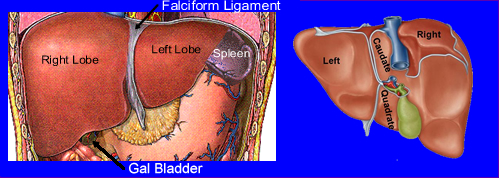
- There are four lobes
- Right and left lobe separated by the falciform ligament
- Two other lobes which lie in the posterior medial portion of the right liver. They are the inferior quadrate lobe located in the inferior portion of the right lobe and the posterior caudate lobe located in the superior portion of the right lobe
- The gallbladder rest in a recess called the gallbladder fossa on the inferior surface of the right lobe of the liver
- Separating the left and right lobe is the falciform ligament that anchors the liver to the diaphragm and the anterior portion of the abdominal wall
- The liver is encapsulated by the visceral peritoneum
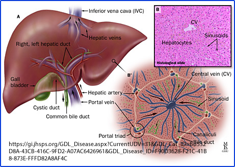
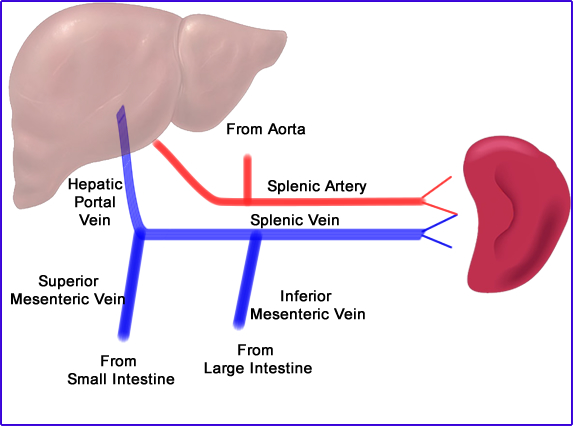
- Blood is supplied by the superior and inferior mesenteric veins that drains into the hepatic portal vein which supply the liver. The hepatic portal vein enters the liver at the portal hepatis along with the hepatic artery. The hepatic portal vein and the hepatic artery then divide into smaller and smaller branches until they finally becoming two parts of the portal triad. Blood from the hepatic portal vein and the hepatic artery which drain into sinusoids becoming the central vein.
- The lobes of the liver have functional units called lobules.
- A lobule is a hexagon cylinder shaped structure
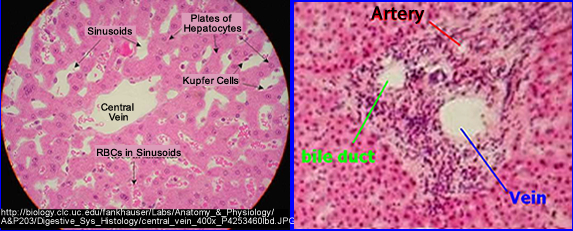
- Each lobule contains specialized cells, hepatocytes and Kupffer cells, with a central vein that supplies blood to the peripheral areas of the lobule through the sinusoids
- Hepatocytes remove nutrients and electrolytes for storage and metabolism
- Kupffer cells, part of the Reticuloendothelial cells (REC), are located along e walls of the sinusoids and phagocytize aging white and red cells, bacteria, and toxic substances
- Around the periphery of the lobule the portal triad contains branches of the hepatic artery, portal vein, and a bile duct
- Blood leaves the sinusoids by draining into the the central vein to the interlobular vein, then the hepatic to the inferior vena cava
- Blood flow
- 75% to 80% of blood flows to the liver comes from venous via the portal vein
- 20% comes from the hepatic artery
- Vascular tumor, such as hepatomas and metastatic disease, may show increased uptake in the arterial phase as seen on a dynamic acquisition
- Why would tumors more likely be seen in the arterial phase?
- Bile is one of the products produced by the hepatocytes and the actual process of bile production will be discussed in a future lecture. For more information you can link to hepatobiliary imaging
- The liver has five major functions
- Secretory - hepatic cells form and secrete approximately 1 liter of bile per day. Bile is made up of bile salts (cholic acid and chenodeoxycholic acid), cholesterol, bile pigments, a variety of electrolytes and water. The bile salts aid in the emulsification of fats in the intestinal phases of digestion. Bile pigments is formed from the hemoglobin of degenerating red blood cells.
- Circulatory - The liver aids in regulation of blood volume by manipulation of the hepatic vein. Erythrocyte-maturing factor is produced which aids in the development of RBCs. The liver also produces coagulation factors: prothrombin and fibrinogen
- Storage of: Vitamins A, D, B12, iron, copper and magnesium and other tracer elements
- Metabolic - The liver synthesizes glycogen, cholesterol, phospholipids, and ketones; and fats from glucose. It also converts glycogen to glucose, and stores glycogen. The liver is a primary organ for fat metabolism. The liver synthesizes proteins, converts amino acids into glucose or fatty acids, and deaminizes (excess amino acids are synthesizing urea)
- Detoxification - The liver detoxifies products of digestion using chemical reactions such as oxidation reduction and conjugation. The liver eliminates drugs in the bile and reduces the toxicity of morphine and strychnine by storing and freeing them slowly. Kupffer’s cells have phagocytic action, which is part of the bodies defense mechanism
When RBCs are broken down: globulin, iron, and bilirubin are released. Bilirubin is the reabsorbed by hepatocytes and excreted, with the end product being bile. It should noted that when the hepatocytes are not functioning correctly then the circulating of bilirubin raises and the patient eventually becomes jaundice.
Return to the beginning of the document
Return to the Table of Content