Clinical Application of PET Imaging of the Brain
This unit contains a lot of information that goes beyond what you really need to know. Therefore, please concentrate on the following:
There is a lot of content in here that goes beyond your need to know, therefore concentrate on the following points:
- Know the details on how to complete a PET brain procedure
- What concerns are there when imaging the brain with FDG - brain uptake vs. disease vs. white and gray matter
- Up/down regulation
- Diseases:
- Application in Epilepsy
- Picks and AD
- Assessment with trauma
- Low vs high metabolic glioblastoma
A discussion of the brain with FDG on-board
- Metabolism
- The use of FDG in the brain can be classified as the assessment of regional metabolic utilization of glucose consumption (rCMR-glu) in brain tissue (1)
- Approximately 25% of the total energy required by the body, occurs in the brain
- Reasons for this highly metabolic rate are:
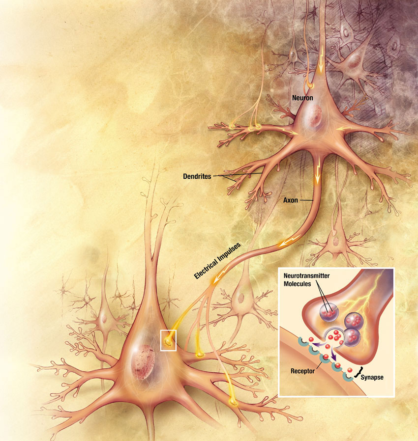
- Need to maintain ionic exchange with the sodium/potassium pump, which occurs with intra/extra cellular components of the brain
- Glucose utilization localizes around the synaptic zones
- Uptake for FDG is greater in the gray vs. white matter, however, uptake in white matter increases if there is a demand to do so
- What is the difference between white and gray matter?
- Grey matter contains your brain function, sometimes referred to as the "living brain." It contains dendrites and axon terminals of the neurons and is the location of the synapses
- White matter is made up of axons that connect the different parts of gray matter
- From a diagnostic point some brain tumors are difficult to see. Reasons?
- FDG uptake may be greater in normal brain tissue when compared to tumor. The "background" hides the diseased tissue
- Smaller lesion with FDG uptake can be missed (based on resolution)
- The greater the metabolic activity of tumor, usually the more aggressive which means greater uptake of FDG
- Other aspects to consider when imaging the brain with FDG
- Excessive eye movement may cause increased uptake
- Any type of outside stimuli may cause "abnormal" uptake
- Muscle and lymphatic uptake
- Resolution is PET is approximately 6 mm
- Imaging Protocol
- Place the patient in a dark quite room
- Establish an IV site at least 15 - 30 minutes prior to the FDG injection
- Inject the FDG without disturbing the patient
- Uptake phase should last 45 - 60 minutes
- The brain should be imaged in only 1 bed position
- The edges of the FOV can slightly distorted an image, for this reason 2 beds are not acceptable
- Setup acquisition for 3D mode and double the acquisition time (MCV protocol) on a single bed
- Use TOF in available
- When placing the head into the scanning consider the following positioning
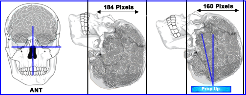
- Use a head holder to restrict movement
- When looking at how the brain/head is position consider the above images
- From the anterior view no rotation or tilt should be seen. You should be able to draw two straight lines that are perpendicular to each other. One line through the nasopharyngeal area and the other through the orbital socket
- Lateral angle, prop up the head up several inches off the table by placing a rolled up cloth underneath the back of the skull. The slight angle improves the area to be scanned. Compare the two lateral images where the tilted brain scan takes up less space
- Why is this important? Sometimes, when you scan an "non-angled" image the volume of the brain will require more than 1 bed position
- If you are imaging the patient because he/she has seizures consider
- Tell the patient not to fall asleep during the uptake phase
- Connect an EEG to the patient to monitor any seizure events
that might occur when he/she is being imaged
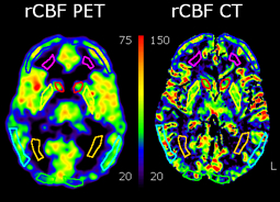 Use of 15O - http://www.ejnmmires.com/content/1/1/28
- rCBF brain imaging with H215O was compared with CT angiography of the brain to determine if CT contrast data mimics rCBF data
- It was concluded that CT data always overestimated rCBF
- That said, a high level of contrast flow may be associated with a positive rCBF rate
- If you are thinking tumor, usuallly there is greater blood flow
- If you are thinking trauma, blood in an area of interest is a positive indication of viable tissue
Diseases of the brain and the associated radiopharmaceuticals
- Infections
- General (FDG)
- The ability to tell the difference between a high grade tumor and infection/abscess is (usually) not a problem, because tumor uptake tends to be more intense (this latter statement requires more research).
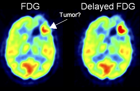
- However, if there is questional infection that displays uptake on the initial scan a delayed image will help determine if it is infection/abscess or tumor
- Another issue occurs with some infections and/or low grade tumors. This relates to the amount of uptake between a low grade tumors and an infectious process, where they may be similar. Furthermore, sometimes the low grade tumor and/or infection, might be masked by the normal high level of FDG uptake in normal brain tissue
- The above image shows questional tumor and then on delays further increase of FDG uptake is an indication that this is a tumors
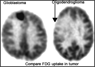
- Here is an example how a high grade glioblastoma obvious has a high level of FDG, whereas a oligodendroglioma has less activity than the surrounding brain tissue
- In addition, there may be difficulty with high-grade tumor that have been treated with radiation
- Uptake that is seen at the edge of the necrotic tissue may contain viable brain tissue or surviving tumor (this could be a hard call)
- Uptake within the necrotic area may contain underlining infection (showing abnormal uptake)
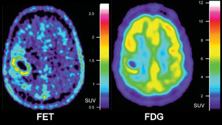
- When FDG uptake is noted another radipharmaceutical is applied, 18Fethyltrosine (FET). Lack of FET uptake is an indication of infection and increased FET uptake is an indication of tumor. Notice how FET has little uptake in normal brain tissue
- Pyogenic abscess
- Infection starts when bacteria arrive at the site of the brain via vascular blood supply. Usually the infection starts between white and gray matter where one or more lesions may develop
- The typical patients with a brain infection may have the following history: drug abuse, underlining sepsis, infection in the sinuses, cardiac infection, or lung infection
- Infections most often are found in the parietal or frontal lobe
- Why is there increased uptake in an infection?
- Stimulation of phagocytosis of white blood cells from an infecious process cause an increase in glucose uptake
- Hexose monophosphate shunt + increase in O2 (can up to x50)
- Refered to as the respiratory burst
- There appeas to be a benefit with acute on chronic levels of infection
- Noninflammatory issues that contain these sames elements do not show increased uptake of FDG
- Toxoplasmosis - T. gondii infects brain tissue and is more typically seen in infants. Uptake of FDG varies between the intensity of the gray and white matter
- Lymphoma vs inflammatory lesion (toxoplasmosis) in AIDS patients where an SUV of greater than 3.8 indicates lymphoma. Therefore PET seems to have an advantages over CT and MRI. Also see See brain cancer.
- Creutzfeldt-Jakob Disease - While CT and MRI have some limited success, FDG seems to show regional decreased uptake in the affected area (more info below)
- General application of PET - infection has an increased uptake in with FDG and there may be issues of infection vs tumor. However, SUV values are higher with tumor/. In addition, over time if two acquisitions are taken, SUV value will drop with infection, but not with cancerous lesion
- Epilepsy
- Overview
- Complex partial seizures usually occur in the medial temporal lobe (MTL). This significant subgroup of patients are classified with temporal lobe epilepsy (TLE). Often associated with TLE is hippocampus sclerosis (HS)
- If the disorder is unilateral then surgical intervention is suggested by removing 2/3 of the temporal lobe on the affected side. FDG should only be used for seizures in the TML. Seizures beyond the temporal lobe 18F-flumazenil FMZ should be administered
- If the disease is bi-lateral surgery cannot be done because it would leave the patient neurologically impaired
- PET imaging is a preferred method to determine if a seizures is caused by TLE and also when the patient is in the interictal state
- TLE is positive when there is reduced activity on the affected side. This can extend laterally into the cortex of the temporal lobe
- The key role FDG is to determine if there is uni or bilateral uptake in the MTL which then determines if surgery is warranted. If hypometabolic uptake is noted beyond the TLE no specific conclusion is drawn
- b-Amino-Butyric Acid (GABA) receptors and the use of FMZ
- Is produced to Inhibits neurotransmission and reduced excitation that causes epilepsy
- Considered a benzodiazepine receptor (BPR)
- GABAA can be imaged with (11C or18F) FMZ
- When compared to FDG, FMZ only shows up in the presence of GABA. Therefore excessive uptake of FMZ is an indication that the brain is attempting to reduce neurogenic activity to inhibit seizures
- FMZ seems to have applications beyond the temporal foci to include: different types of neocortical focal epilepsy, parietal and occipital lobe epilepsy
- Several articles discuss the use of FMZ to determine partial seizures of the neocortex (2)
- Other tracers
- 11C-carfentanil, 11C-diprenorphine, 18F-cyclofoxy will attach to certain types of opioid receptors in the brain. It is believed that some opioids receptors reduce or limit the area of seizure foci, as in the cast where : opioid receptor show increased uptake with 11C-carfentanil
- Another example11C-deuterium deprenyl shows increased binding to monoamine oxidase, an enzyme found which is found HS tissue
- Ictal
- PET is generally not useful in this area simply for the fact that it has such a short half-life (when compared to 99mTc)
- Therefore, ECD or HMPAO are the preferred agents since the radiotracer can be injected when the patient goes into a epileptic state (time is the key factor)
- General application of PET is to define MTL in the interical state, but not in the ical state
- Dementia
- General comment (two types)
- First type of dementia is directly associated with cognitive behavior, with no other underlining pathology
- Second type of dementia occurs because there is some other underlining pathology (ex. brain tumor or infection)
- Alzheimer's disease (AD) [3]
- Disease
- This neurological disorder progresses over time and may occur anywhere between the ages of 40 to 90 years
- AD is believed to be an inflammation process, specifically related to beta-protein fragments from the beta-amyloid precursor (glycoprotein). This fragments forms amyloid plaques resulting in neurofibrillary tangles, in turn, this regression slowly causes the brain to atrophy
- Imaging
- FDG-PET is sometimes difficult to image in its earlier stages (~ 66% sensitive). In time, even the false negative patients become positive
- Cortical atrophy starts with decreased uptake in the temporal and parietal lobes and then usually progresses to the frontal lobe. In addition, enlargement of the 3rd/4th ventricles and the choroids-hippocampal fissure are noted
- The earlier you treat the disease with cholinesterase inhibitors the better the patient will respond. Therefore, early intervention is critical
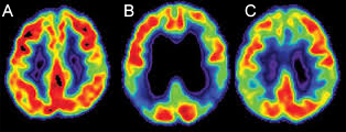
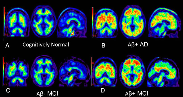
- The above images demonstrate: A - Normal, B - Mild/moderate AD, and C - More advanced AD
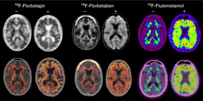
- Radiopharmaceuticals
- FDG is the most common tracer used
- , Flutemetamol, Florbetaben, and Florbetapir have been approved for routine use however there is currently limited reibursement from insurance companies
- Each agent works in a similar fashion
- Cross the BBB and localize on to amyloid-β (Aβ) protein
- Also associated with Alzheimer's and Aβ are tangles of tau protein
- Sometimes patients may show both Aβ and tangles, but are asymptomic
- Florbetapir, Florbetaben, and Flutemetamol all bind to amyloid plaque and appear to be a positive indicated of AD
- However, in a resent article by Chiotis K, et al. (2020) it appears that [18F]THK5317 may even have greater sensitivity in identifying AD as it binds to neurofibrillary tangles, tau protein. When compared to [11C]PIB and 18FDG
- The difference was 95% percision vs 77% percision
http://www.sciencedirect.com/science/article/pii/S2253808915000579
- Comparison and contrast all three above
http://medicalphysicsweb.org/cws/article/research/50285
- Above - A is normal, B - is AD positive with with amyloid, C - mild cognitive problems but no amyloid plaque, and D - patient that progressed into dementia.
- Procedure differe slightly between the agents (more information about a specific agent click the link below)
- Florbetapir (AmyvidTM) - Dose is 10 mCi, delayed images start 20 to 50 minuts post dose, acquire data for 10 minutes on a single bed
- Florbetaben (NeuraceqTM) - 8 mCi dose, delayed images start 90 post dose, acquire single bed position for 20 minutes
- Flutmetamol (VizamylTM) - 5 mCi dose, delayed image starts 45 to 180 minutes post dose, acquire a single bed position for 15 to 20 minutes
- Multi-infarct Dementia (MID)
- Disease
- Second most common dementia
- Asymmetrical infarcts that spreads throughout the cortex and occur within a limited time frame (unlike AD)
- Imaging
- CT and MRI are appropriate for this type of diagnosis
- PET shows reduced uptake in the affected areas with MRI correlation. However, FDG-PET tends to show greater involvement when compared to MRI and has some purpose for imaging this disease
- Frontotemporal Dementia (FTD) - originally referred to as Pick's disease
- Disease
- This neurodegenerative disease causes atrophy of the frontal and temporal lobes
- In Pick disease pick bodies are present in histological analysis
- Imaging
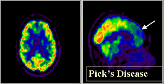
- Looks similar to AD, however, in Pick's hypoperfusion is noted in both frontal and anterior temporal lobes
- CT and MRI may show an abnormal increase in the interhemispheric fissure and enlarged frontal horns
- Normal-Pressure Hydrocephalus
- Disease shows normal CSF pressure within dilation of the 3rd/4th ventricles. CSF shunt is used to treat this disease. The patient may be presented with a triad of issues: dementia, urinary incontinence, and ataxia (uncoordinated muscle movement)
- Usually diagnosed with CT or MRI. In PET-FDG reduced perfusion is seen on a global scale and ventricles should appear enlarged
- Alcohol-related dementia
- There is an apparent decrease in uptake in a global scale or at the regional level, sub/cortex or even the left hemisphere
- Reduced uptake has been seen in chronic alcoholics and specifically in the areas of occipital, prefrontal, and cerebella cortices. Furthermore, these areas have been known to contain high level of GABAA receptors
- Creutzfeld-Jakob disease (CDJ)
- Disease caused by a (proteinaceous infectious particles) prion. Spreads quickly through the brain creating a spongiform type tissue. In a very short time the patient goes from dementia to death
- Decreased uptake with PET-FDG is noted, however, actual clinical data is limited
- See example of CDJ
- AIDS-related dementia
- HIV involvement in the brain or a result of an opportunistic infection (toxoplasmosis) or AIDS-associated neoplasm
- Approximately 5% of AIDs patients have this related dementia
- PET-FDG shows hypoperfusion in the cerebral cortex
- Extrapyramidal disorders (ED)
- General comments
- These are different types of movement disorders
- Disease cause patients to appear rigid, have nervous tremors, show reduced movement (bradykinesia), bad (degenerative) posture.
- CT and MRI may show disease, however, PET usually picks up the disease(s) at an earlier stage
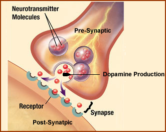
- At issue is the regulation of dopamine at the synapse, which transmits an electrical pulse from one neuron to the next
- If dopamine is being over produced receptors sites decrease causing "down regulation"
- If dopamine is lacking then the response is to increase the amount of receptors resulting in "up regulation"
| RAC Uptake |
FDOPA Uptake |
Regulation |
DOPA Production |
Receptor Sites |
| Increase |
Decrease |
Up |
Decrease |
Increase |
| Decrease |
Increase |
Down |
Increase |
Decrease |
- Uptake of FDOPA depends on the rate of Dopamine produced
- RAC Uptake relates to the the number of D2 receptors
- The above chart further illustrates this concept with FDOPA (demonstrates the amount of dopamine production) and RAC (illustrates the amount of D2 receptor sites
- Sometimes there is degeneration of the entire system
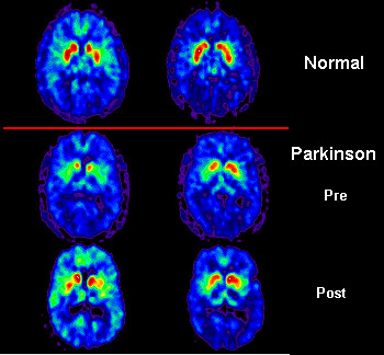
- Parkinson disease (PD)
- Disease
- Nonindustrial projections to the basal ganglia degenerate where there is involvement of the putamen, but not the caudate
- Degeneration occurs at the pre-synaptic side of the dopamine system while the post synaptic remains intact with increased receptor sites. This causes a loss in dopamine production and requires dopamine therapy (supplement)
- Up regulation is noted
- Imaging
- FDG uptake is excessive which may be do to the increased firing of the basal ganglia that is caused by the lack of dopamine. Because of this FDG is not recommended. Why?
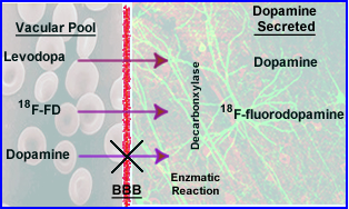
- 18F-fluorodopa (PD) evaluates the dopamine pathway and indication the lack of radiotracer where . Related to dopamine production
- Comment on dopamine production: (1) While FD crosses the BBB, dopamine cannot (because dopamine has a charge preventing it from crossing), (2) However, FD and Levodopa do cross the BBB via enzymatic reaction of decarboxylase, (3) these chemicals converts to "dopamine"
http://www.cerebromente.org.br/n01/pet/pet.htm
- The above images show uptake of FDOPA in: (1) normal brain, (2) reduced uptake in the putamen, (3) improved uptake to the affected area following dopaminergic therapy
- 11C-raclopride (RAC) evaluates the amount of the amount of D2 receptors and will correlate to an increase in uptake. Most movement disorders, with the exception of PD, show a reduction in D2 receptors
- Multiple system atrophy (MSA)
- Disease
- Neurodegeneration in nature and occurs in 10% of the ED population
- Involves gliosis in the basal ganglia, brain stem, cerebellum, and spinal cord
- Pre/post synaptic degeneration occurs at the putamen and caudate
- Imaging
- PET -FDG shows reduced uptake in the striatum and cortical areas
- FD and RAC show reduced uptake
- Dopa-responsive Dystonia
- Disease
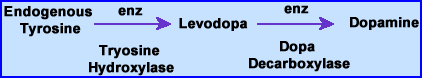
- Rare genetic disorder where the body does not synthesis tyrosine hydroxylase, the prevents the production of dopamine.
- The process is noted above where endongenous tyrosine converts to Levodopa to dopamine
- It is first seen in childhood with dystonic (abnormal movement or muscle tone) posturing and then as a adult the condition worsens
- Imaging
- Sometimes reveals a slight loss in nigrostriatal dopaminergic neurons
- Some up-regulation occurs with the D2 receptors
- There is a positive response to the therapeutic administration of Levodopa
- Corticobasal degeneration (CBD)10
- Disease
- Sub/cortical atrophy with movement and memory problems
- Frontoparietal, cortex, striatum, and thalamus can be effected
- Imaging
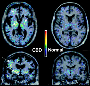
- 11C(R)-PK11195 is a biomarker of sites that express benzodiazepine where there is an increase in microglial cells found in the one or more areas of the: caudate nucleus, patamen, pons, pallidum, and substrantia nigra
- Reduced fluorodopa is seen in the caudate and the putamen (~25%)
- 123I-IBZM shows reduced binding in the D2 receptor
- Huntington disease (HD)
- Disease
- Genetic issue with significant loss of motor movements occurs over time
- Cognitive issues occur between 30 -40 years of age with neuron deterioration and gliosis within the striatum
- Microglial activation may play an important role in neuronal death, since excessive amount has been found on autopsy (6)
- Imaging
- Caudate nucleus and putamen show decreased FDG uptake
- The greater the reduction of uptake usually the greater the dementia. Frontal, parietal, and temporal lobes have been effected
- CT and MRI find atrophy, however, this occurs later
- Progressive Supranuclear Palsy (PSP)
- Disease
- PD type symptoms with vertical eye movement that occurs between the ages 60 - 70
- Cell loss is noted in: brainstem, pallidus, and substantia nigra (no complex involvement)
- Midbrain atrophy occurs
- Imaging
- FDG uptake is reduced in the: superior frontal cortex, basal ganglia, thalamus, and pons
- Pre-post synaptic areas are reduced with the putamen being affected
- MRI shows enlargement of ventricles
Assessment of cerebrovasulcar disease
- Disease
- Transient ischemic attack (TIA)
- Stenosis within the arterial areas of the brain will causes hemodynamic changes
- Atherosclerotic plaques reduce blood flow. Plaques can break off which may cause a stroke
- Alterations of blood flow (reduced) for whatever reason can lead to more severe issues and must be treated
- How does the brain respond to this reduced perfusion?
- Initially an attempt to increase the extraction fraction of oxygen
- Secondly, dilation of the involved artery(ies)/arterioles
- Over time, as the condition worsens the patient will become symptomatic: loss of vision, alteration in speaking (aphasia), weakness or numbness involving one side of the body
- Moyamoya disease (means "something hazy just like a puff of cigarette smoke drifting in the air")
- Abnormal blood flow occurs at the base of the brain
- Stenosis, occlusions, and collateral circulation occurs
- Usually in the areas of the internal carotid arteries
- Depending on age
- Younger patients tend to exhibit TIA type symptoms
- Older patients tend to have intracranial bleeds
- Acute ischemic stroke
- Consequences are usually severe, however, literature indicates that if treated early, re-vascularization may salvage some of the "damaged" area
- Zones of a stroke
- No intervention will help the necrotic tissue caused by perfusion loss
- Surrounding the stroke zone is an area referred to as the penumbra
- If re-vascularization occurs the penumbra tissue may survive (consider the cardiac as another example)
- PET data indicates that tissue with <12 mL/min/100g cannot be salvaged
- PET data that is between 12 to 25 mL/min/100g in the penumbra are salvageable pending re-vascularization and how soon this is done post infarct
- Any tissue that is <65 mL/min/100g and lacks any intervention will eventually become necrotic (time frame - no more than 2 weeks)
- 11C-Flumazenil has been shown to useful to evaluating the penumbra via the uptake of this radiotracer as a candidate for re-vascularization
- Assessment of disease
- The terms
- rate of cerebral blood flow - rCBF
- rate of cerebral blood volume - rCBV
- [15O]CO is strictly bound to hemoglobin
- Hence its concentration in the brain determine the amount of blood in the brain
- Cerebral perfusion reserve - PR
- Evaluation of disease and its related quantification
- The ratio of CBV to CBF is essential in determining if ischemia is present
- Initially a base line scan completed with [15O]H2O
- A second scan is acquired, but only after 1 g of acetazolamide (causes vasodilation)
- Administration of the vasodilator increases CBF. Administration of CO2 will cause the same affect, a hypercapnia state
- The ratio of the hypercapnia state over baseline expresses PR
- If a vessel is stenotic on one side of the brain, how does this compare to non-affect side?
- The body response to chronic vascular disease is to vasodilate the affected side
- Hence, a baseline exam would reveal normal blood flow to both areas
- If the patient is then put into a hypercapnia state or given acetazolamide, then increased blood flow result to the unaffected side (consider coronary steal with the heart)
- Hence, a PET scan would reflect reduced uptake to the affected side and the comparison to the baseline study
- Quantification standpoint - Activity collected for 60 seconds was normalized to the injection per kilogram of body weight (Activity/kg). This was compared to 8 different regions of the brain to evaluation rCBF. Linear correlation determined the baseline
- Treatment
- Re-vascularization
- Endarterectomy (removes plaque from inside the artery) or stenting or intra/extracranial bypass surgery
- Consider the similarities when treating heart disease
Cancers of the brain
- A look at the normal brain
- Brain utilization of glucose directly correlates to the amount of FDG uptake
- Greatest amount of glucose consumption occurs with the brain
- Since gray matter is more metabolically active than white matter, FDG uptake is greater in gray matter. Normal ratio is 2.5 to 1.0 (gray to white)
- Other areas around the brain also pickup FDG and can falsely influence a diagnosis in this region. These areas include: muscles, glands, and lymph
- Brain activation studies can be done where a specific stimuli (e.g. talking, hearing, visualizing) an be analyzed with PET
- Imaging the brain with FDG-PET
- Best resolution for PET is around 6 mm
- Specific anatomical areas that can be seen with PET include: brainstem, thalamus, cerebellum, head of the caudate, putamen, and cortex
- MRI and CT (with contrast) should be used in conjunction with PET for better anatomical analysis
- Because of the excessive amount of activity in a brain lesions that may be similar to the amount of uptake to gray matter, this type of tumor can be missed. Consider target to background, where your background activity is very high. Co-registration with MRI or CT will reduce this type of false-negatives
- PET-FDG procedure
- NPO for at least 4 hours
- Setup patient's IV prior injection
- Place the patient in a quiet room that has little to no lighting
- After explaining the procedure and taking the patient's history wait at least 15 minutes before imaging
- Tell the patient to keep his/her eyes open after injection
- Patient can be imaged 30 to 60 minutes post dose
- It is suggested that any abnormality found on PET should be further documented with MRI
- Primary brain tumors
- Imaging the brain
- The use of MRI over CT is the preferred method for evaluating brain tumors
- PET can be used to diagnosis, define the level of malignancy (low, moderate, high) based on degree of uptake (FDG), used to assist in guiding a biopsy, determine survival, monitory and assess therapy, and determine viable tumor/necrotic tissue
- At issue with PET is the excessively high level of normal FDG in brain. Its greatest concern is tumor uptake in the area of gray matter, because gray matter extracts higher levels of glucose (hot normal tissue = activity from viable tumor)
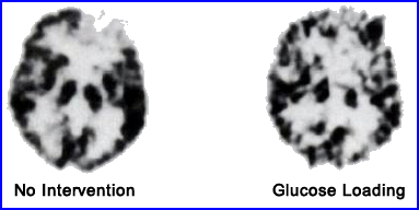
- Ishizu, K, et al. suggest the utilization of glucose loading to improve tumor uptake. Administration of IV glucose and FDG were administered at the same time causing a reduction of FDG uptake in brain tissue and tumor. However, because there was significantly less reduction of tumor uptake, the tumor-to-cortex ration actually increased, causing improved tumor visualization7
- Primary brain tumors
- In children this is the second highest form of cancer, while in adults it is considered rare (11 - 12 per 1000,000)
- Gliomas comprise approximately 51% of all primary brain tumors
- Glioblastoma (multiforme) is the most common type seen with the average age of onset at 62 years
- Glioblastomas arise from glial (supportive) tissue within the brain
- Astrocytic and oligodendroglioma tumors arise from epithelial cells and include the following tumor types: astrocytoma, anaplastic astrocytoma, glioblastoma, gliomorphic xanthroastrocytoma)
- A chart is provided on the different types of brain tumors, its tissue type, and rs that occur in the brain
&
- Diagnosing and evaluation primary brain tumors with PET
- Tumor uptake
- In general, the greater the amount of FDG uptake the more aggressive the cancer and the shorter the survival time for the patient
- Low grade tumors have similar to less activity when compared to white matter while high grade tumors may have activity equal to or even greater than that of gray matter (tumor is compared the contralateral side of the brain)
- Tumor to white matter ratio of 1.5 (or more) and tumor to gray matter ration of 0.6 or more where classified as high grade
- High grade tumors usually have heterogeneous uptake
- Biopsy - Since high grade gliomas are usually heterogeneous, guided biopsy with PET is suggested. Why? If the biopsy collected tissue that had reduced uptake, the tumor might be underscored
- Prognosis
- Patients that had a low grade tumor had a 1-year survival rate of 78%
- Patients with a high grade tumor had a 1-year survival rate of 29%
- Therapy results
- Following surgical resection PET can differentiate viable tumor from surgical scarring
- In radiation treatment reduced FDG is noted within necrotic areas of the tumor while viable tumor will pickup the radiotracer
- High dose radiation initially shows increase in uptake of FDG which may be due to increased macrophage activity and is not related to tumor viability
- Following stereotactic surgery reoccurring tumor can be detected up 86% of the time
- Steroid therapy does effect FDG uptake in white/gray matter, but does not alter FDG uptake within a viable tumor
- Seizure foci has reported near sites of therapy causing false related increase to FDG levels
- Other PET radiopharmaceuticals
used in the brain
- Amino Acid metabolism
- 11[C]methionine
- Via breakdown of the BBB or by increased amino acid metabolism within the tumor may relate to the tumor viability and increased uptake
- Brain tissue has little to no uptake of these amino acids, therefore giving this PET agent greatly improves target to background when compared to FDG or FET
- 11[C]choline utilization with lipid metabolism
- Cancerous cells have increases levels of phospholipids that can be evaluated with primary brain tumors and prostate cancers
- 18F choline may also be viable
- Oxygen
- 15O inhaled evaluates oxygen uptake with tumor cells. Higher rates of oxygen exchange relate to longer survival rates
- H15O2 has been used on patients for evaluation prior to surgery
Other types of brain cancer
- Diagnosing other types of brain
cancer
- Types of cancers include: metastatic disease (primary is outside the brain), intracranial lymphoma, and nongliomatous primary brain tumors
- MRI is the method of choice for diagnosing disease in the CNS
- PET-FDG may be limited in this arena
- What causes uptake of a radiopharmaceutical
- Vascular follow to the disease process
- Ability for the area of interest to pickup the radio-tracer
- Agent crosses the BBB
- Tissue binding via specific cells or involvement into some type of metabolic pathway
- Metastatic disease
- FDG has poor contrast considering the high background counts contributed by the gray matter. Therefore lesion detection may be difficult to diagnose
- Problem - If metastatic brain disease is missed then an improper therapeutic response will be given to the patient
- Other PET agents should be considered
- 11[C]methionine - as mentioned earlier. Chang et al. evaluated 45 patients and found four of five metastatic brain lesions. FDG either missed it or these lesions had only slight FDG uptake
- 11[C]Choline - mentioned earlier. Pietermann et. al. studied 17 patients with brain mets and found 23 lesions with choline where FDG only diagnosed 3
- On the reverse side - FDG was able to find the unknown primary cancer that had metastatic lesion in the brain 82% to 100% of the time. These primaries found included: lung, breast, melanoma, and GI track tumors
- Primary intracerebral lymphoma
- According to study done by Karantanis D, et al. FDG is useful in cranial (87%) and spinal cord (80%), but not with ocular involvement (20%) in patient that did not have HIV7
- In HIV patients PET-FDG can differentiate between tumor and infection 80% to 95%
- PET may show be applied to therapy monitoring and biopsy, however data is limited
- Nongliomatours primary brain tumors
- FDG
- Uptake has been noted with Meningiomas and schwannomas
- While pituitary adenomas are usually benign in nature there is apparently significant FDG uptake
- Reduced uptake 11[C]Choline following therapy is considered a positive prognosis
- More research needs to be done in this area to determine the benefits of the different PET radiopharmaceuticals in the areas of detection, treatment evaluation, and reoccurrence of disease
Return to the Table of Content
References
1 - Neuronuclear Assessment of Patients With Epilepsy. Goffin, K., et al. Seminars in Nuclear Medicine (2008)
2 - Gray and white matter FMZ binding in neocortical epilepsy with normal MRI. A PET study of 44 patients. Hammers A, et al. Brain (2003)
3 - The Significance of Neuroinflammation in Understanding Alzheimer's Disease. Eikelenboom, P, et al, Jour Neural Transmission (2006)
4 - PET Imaging of the Peripheral Benzodiazepine Receptor: Monitoring Disease Progression and Therapy Response in Neurodegenerative Disorders. Doorduim J, et al, Current Pharm Design (2008).
5 - Early [11C]Flumazenil/H2O Positron Emission Tomography Predicts Irreversible Ischemic Cortical Damage in Stroke Patients Receiving Acute Thrombolytic Therapy. Heiss, WD Stroke (2006) .
6 -
Microglial activation in presymptomatic Huntington's disease gene carriers. Tai, YF, et al Brain, (2007)
7 - Effects of Hyperglycemia on FDG Uptake in
Human Brain and Glioma. Ishizu K, et al. (1994)
8 - 18F-FET PET Differentiation of Ring-Enhancing Brain Lesions. Floeth FW, et al. (2006)
9 - Avid amyloid tracer hits target in Alzheimer's study, Reuters, 6/11/2010
10 - In vivo imaging of microglial activation with [11C](R)-PK11195 PET in corticobasal degeneration. Gerhard A, et al. (2004)
11 - Babior BM. The respiratory burst of phagocytese. J Clin Invest. 1984;73(3):599-601.
12. Chiotis K, Savitcheva I, et al. [18FTHK5317 imaging as a tool for predicting pospective conitive decline in Alzheimer's disease. Molecular Psychiatry (2020)
FET- Is an amino acid analog. Uptake of these agent have several characteristics: (1) it is independent of vascular flow, (2) does not require the breakdown of the BBB. (3) But does demonstrate upregulation of amino acid transport, which occurs in tumors. This type of uptake can be seen in both low and high grade gliomas. In contrast, normal brain tissue does not metabolize amino acid. Results = improved target to background
7/20