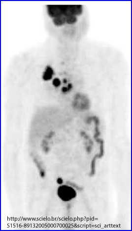
This 82 year old male was initially being staged for lung cancer and was imaged with FDG. His PET scan not only found the lung cancer, but also found disease in the colon. A colonoscopy found a 3cm tubulovillous adenoma in the area of the sigmoid colon. Another tubulovillous adenoma was discovered in the cecum.
In the above image a lot of increased uptake is see with FDG. Of those hot spot/areas, which is/are disease and which displays normal FDG uptake.15