Image Registration in the presence of large geometric changes in the lung
A variety of tasks in radiation therapy require image registration – the process of identifying the locations of corresponding anatomy on different images. Some of these include target localization for radiation delivery, adaptive radiotherapy, and assessment of cumulative delivered dose. Our lab’s role in this area has been to design and implement novel registration algorithms designed for specific tasks. In one example, we designed an algorithm that could robustly and quickly register tumors of changing shape and size for image-guided radiation therapy. We are also developing deformable image registration to handle challenging cases of large changes during therapy (for example, partial lung collapse in one image but not in the other).
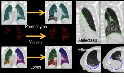
Mass-Preserving Image Registration
Atelectasis is the partial collapse of lung tissue, which often accompanies lung tumor presence. The goal of this sub-project is to adapt mass-preserving image registration, which was previously developed to account for lung density changes during free breathing, to register pre- to post-atelectasis CT.
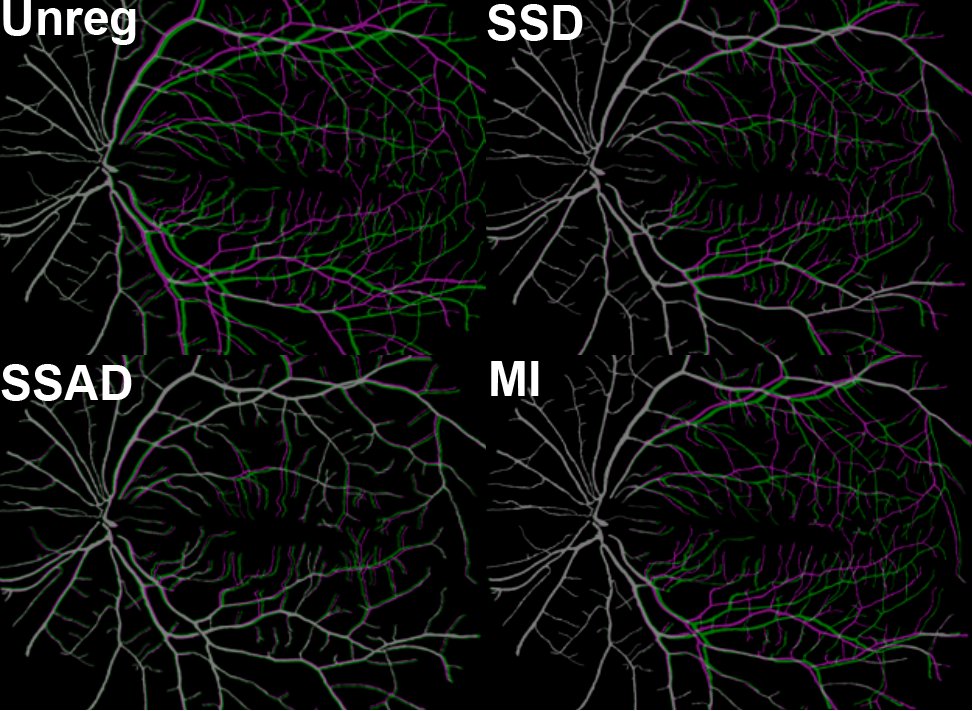
‘Fuzzy’ Registration of Pulmonary Vasculature
Registration of lung tissue under large geometric changes is challenged by the lack of robustness. Registrations often match vessel branches incorrectly, as they tend to look similar on a local basis. Here, we extend the concept of local histograms to a 2D histogram approach to match enhanced vessel images to improve the robustness of the matching. The following image shows a comparison in 2D test data of conventional (SSD, MI) metrics vs. our proposed sum of squared shape attribute difference (SSAD) metric.
Most registration algorithms used in radiotherapy are based on the small deformation (Demons, b-spline, etc.) model. Varifolds uses a large deformation model and a ‘fuzzy matching’ approach to account for large deformations, and match extracted vessel segments and lung surfaces. Our ultimate goal is to combine varifolds with shape attributes, and incorporate into the registration framework.
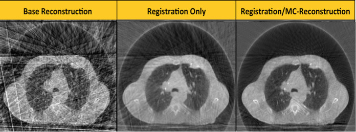
Motion-Compensated Cone Beam CT Reconstruction
Motion during CT and cone beam CT acquisition induces blurring and artifacts that can impact image quality and reduce the visibility of the target and organs at risk. Motion-compensated CBCT reconstruction was proposed by others several years ago, but generally requires a motion model to be built from diagnostic 4DCT data. When the anatomy changes during radiotherapy, this model may no longer be valid. Here, we propose to use a groupwise registration approach to build a model directly from the CBCT data, and then employ this model to perform a motion-compensated CBCT reconstruction.
This project is funded by the National Institutes of Health, Award No. R01CA166119.