Radiopharmaceutical Characteristics and Their Physiological Applications
- 11Carbon =
- Properties
- Synthesis

- T1/2 = 20 minutes
- Its short T1/2 requires the use of an in-house cyclotron
- 11C-acetate =
- Can be used to image the heart, prostate cancer, and Hepatocellular carcinoma
- In general it measures oxidative metabolism
- Proteint synthesis
- Is the smallest of all fatty acid
- It is eliminated by the release the of 11C20
- Prostate Imaging and hepatocellular carcinoma
- In theory acetate uptake will occur in metabolically active (rapid growth) tumor cells that that with aa low oxygen environment and have a high lipid pool
- Uptake may also relate to the testosterone hormone and it relation to the cancer's growth
- The effectiveness of imaging prostate disease is still under investigation, but application of the positron radiopharmaceutical may help define the extent of and determine treatment options (ex. level of surgery vs. radiation)
- Interesting point - because elimination occurs by the exhalation of 11CO2no urinary activity is noted. Such is not the case, if one where to evaluate the prostate bed with FDG
- Application in hepatocellular carcinoma
- As with prostate imaging uptake is associated with increased lipids within the tumor site and its rate of cell growth
- Additionally, there is no hepatocellular excretion of carbon though the hepatobiliary tree, therefore enhancing target to background
- Cardiac Imaging
- Uptake is identified when blood flow reaches the myocardial cells and viable myocardial cells extract the acetate via oxidation metabolism with a by-product of 11CO2
- Reduced or lack of uptake is an indication that there is poor or no oxygen turnover in myocardial tissue
- Therefore, 11C-acetate evaluates oxygen utilization
- This tracer has been used to assess patients with stable chronic coronary disease or post myocardial infarction
- 18FDG and 11C-acetate can be combined to evaluate myocardial ischemia. In this process 11C-acetate has reduced uptake with ischemic cells because of the low level of oxygen. (oxidative metabolism requires a aerobic environment). 18FDG demonstrate increased glucose utilization (and uptake) because uptake occurs with anaerobic glycolysis. Lack of uptake with both agents defines an infarct
- Similar physiologic finding can be noted with 11C-pamitate =
- Comments on imaging
- Can be done in the dynamic mode or as a 5 minute perfusion delay
- In myocardial imaging it is used with 15O for perfusion to evaluate myocardial perfusion
- 11C-glucose
- There may be some application to brain and cardiac imaging
- Current literature shows limited application of this radiopharmaceutical with only a total of 5 written articles within the last 10 years
- 11C-methylspiperone
- Used to assess neurological disorders
- Has a high affinity of D2 receptors
- 11C-methionine
- This amino acid is used to image glial tumors and pituitary adenomas
- Certain tumors, such as glial, have higher concentrations of amino acids
- Uptake is seen as a ratio where the concentration of the radionuclide within the tumor is compared to its concentration in health tissue.
- In addition, the amount of tracer uptake is indicative of low vs. high-grade tumor
- 11C-choline =
- Has an affinity for certain cancers: prostate, colon, bladder, hepatocellular, and gynecologic
- This tracer incorporates into the tumor cell membranes that are composed of phospholipids and specifically phosphatidylcholine where choline becomes an component of the cell membrane
- Looks at the uptake of fatty acid metabolism than analyzes the rate of transport across the cell membrane
- Does not excrete through the urinary system and therefore enhances target to background in the prostate bed and its associated lymph node
- Imaging
- On a fasting patient
- injected and acquire a 45 minute delay
- 11C-palmitate (3 and 4) =
- See 11C-acetate and note that the main difference is size
- Literature mainly focused on the myocardium
- If the heart's in a fasting state its oxidative metabolism extracts fatty acid for energy
- In the presences of excessive glucose, palmitate uptake will be reduced and/or washout rate will increase
- Palmitate is a long change fatty acid
- Approximately 70 - 90% is immediately utilized with the <30% stored in the triglyceride pool
- There are two uptake phases (early and late) referred to as bi-exponential
- Early phase, 50% extraction fraction, relates to oxygen consumption in which beta-oxidation cause palmitate to form 11C-acyl-CoA and release of 11CO2 within tricarboxylic acid (TCA) cycle of the mitochondria
- Second phase relates to the its incorporation triglyceride and phospholipids. The involvement of these exogenous fatty acids apparently complicates the analyze of data. For this reason 11C-acetate is preferred since there seems to have little to no involvement of exogenous fatty acid uptake. Consider acetate shorter fatty acid chain and how this could enhance physiological uptake
- For myocardial assessment refer to 11C-acetate
- Apparently this radiotracer can also be used to assess the oxidative state of the brain where 50 - 60% of its oxidation will produce 11CO2
- The frustration of evaluating this radiotracer is simple -- there are too many animal studies and not a lot of human discussion
- Image - 5 minutes post injection
- 18Fluorine =
- Properties

- T1/2 = 110 minutes
- 18F - NaF =
- Is a salt
- Goes to calcium hydroxyapatite and exchanges with (PO4-)hydroxyl group
- Currently not being used as a bone imaging agent
- Image - 1 to 2 hours post injection
- 18FDG =

- FDG as a "fake" sugar
- As noted above, fluorine replaces one of the hydroxyl groups on the glucose carbon ring
- Metabolically active tissue within the human body (normal tissue/disease) that requires glucose will pickup and initially metabolized FDG
. Within the cell it phosphorylates by adding a phosphate to the ring by hexokinase. It becomes FDG (or) glucose-6-phosphate. However, since flourine has takn e over an OH spot this metabolic process stops. Dephosphorylation occurs, glycolysis stops, and the FDG gets looked into the cell.
- If radioactive glucose had been labeled it would eventually leave the cell, therefore, it wouldn't be a good imaging agent
- Hence metabolically active areas of the body can imaged
- Normal uptake is seen
- Brain - only uses glucose
- Nasopharyngeal
- Myocardium uses both fatty acid and glucose and therefore uptake depends on the state of the myocardium
- Muscle uptake depend on increased activity
- Liver, spleen, reproductive
- Kidney/bladder is seen since FDG is excreted (unlike glucose)
- Inflammation does pickup FDG
- Uptake to scan - consider tumor, tissue and inflammatory uptake, tumor increases over the other at about 45 minutes, but for best target to background a 90 minutes post injection is recommended
- Oncology
- At present the following cancers are being imaged with this radiopharmaceutical: breast, colorectal, esophageal, head and neck, lung (non-small cell), lymphoma, melanoma, single pulmonary nodule, and thyroid cancer
- Imaging cancer may be done for: initial diagnosis, staging, restaging, pre-chemo/radiation therapy, during/post chemo/radiation therapy, and pre-surgical planning
- Neurology
- Seizure evaluation prior to surgical intervention
- Mental disorders
- Tumor viability
- Cardiac
- Primarily done for myocardial viability study
- Not done to analyze ischemia vs. infarct
- Imaging
- Oncology - inject and image 60 - 120 minutes post dose, where max uptake occurs at 45 minutes
- Brain - Setup IV prior to injecting and waite 15 minutes before injection. Delay can occur between 30-60 minutes
- Cardiac - Elevated sugar level, inject once it starts to drop. Then scan 30 minutes post dose
- 18Fluoroethylcholine
- Biodistribution
- 18F-FECh enters tumor cells by active transport
- Via phosphorylation it becomes phosphoryl-18F-FECh which in turn becomes a phospholipid
- This agent has a high avidity for prostate cancer and has similar biodistribuiton prosperities seen with 11C-Choline. There were two differences
- FECh was excreted significantly faster then Choline
- Spatial resolution was better with the fluorine tracer which may relate to the distance traveled by the beta particle
- 18Fluorothymidine (FLT) markers of proliferation (5) =
- Detects amino acid synthesis at the cellular level, which occurs in the DNA. FLT becomes trapped inside the cell via phosphorylation by
thymidine kinase-1 (which is an enzyme) or high cellular proliferation. Thymidine kinase-1 is seen in high concentrations where there is significant cellular growth, ie. malignant cells. Uptake has been seen in colorectal/lung cancers
- Unlike FDG, It does not accumulate in inflammatory lesions
- Can be used for diagnosis of disease and evaluation following therapy
- Production - yeild is very poor: 8 - 14%
- Imaging
- Dynamic and static (1 hr) can be completed
- Biodistribution: bone uptake, hepatic uptake, no brain uptake, no GI uptake
- Brain tumor
- 18F-Fluoromisonidazole (FMISO) (6) =
- FMISO is a derivative of nitroimidzole (type of antibiotic) and is trapped within cells that are hypoxic
- Understanding the physiology of tumor growth
- Often blood flow cannot keep up with excessive tumor growth, which results in a hypoxic state
- In addition, it is believed that tumor progression within the hypoxic state makes these cells more resistant to radiation/chemotherapy
- Can also be used for the heart in viability
- FMISO is found exclusively in hypoxic areas at 1 - 5 minutes post injection.
- High levels have been found in associated high grade gliomas, but not in low grade
- Imaging several hours out is not recommended because data becomes inconclusive
- May used for monitoring and directing hypoxic therapy
- Imaging - Can be done dynamic or static, but 10 to 60 minutes post dose is acceptable
- 18F Flourodopa (F-Dopa)=
- Usually used to image Parkinson's disease and apparently a good imaging agent for neuroendocrine tumors
- In Parkinson's disease, the receptor sites do not function well and F-Dopa becomes flourodopamine via aromatic amino acid decarboxylkase (AAAD). Because there is degeneration on the pre-synaptic side (post is normal) FD cannot be converted to flourodopamine resulting in decreased uptake in the effected area
- Must pretreat with carbidopa to inhibit metabolites (60 to 90 pre-injection)
- Imaging - Usually done dynamic
- 13NH3 =
- Properties
- T1/2 - 10 minutes
- Primarily used to image the myocardium
- Has been shown to have some utilization in brain imaging
- It's short T1/2 requires the use of an in-house cyclotron
- 13N-aminona as s Cardiac agent
- Primarily uptake occurs via blood flow to the myocardium, however, as flow rates increase uptake plateaus
- Uptake of 13NH3
- In the vascular pool this agent is mostly in its ionic form 13NH4
- When extracted it converts back to 13NH3 which makes it lipid soluble, crossing the cell membrane of myocytes
- Once trapped to it encounters glutaminic acid and via an enzymatic reaction becomes13N-glutamine
- This radio-agent is recommended for myocardial viability. The question is why? This may be the answer
- Hibernating myocardium is very ischemic and has an anaerobic environment
- As long as there is some form of blood flow then13NH3 will be picked up by the viable tissue. It is then synthesize to [13N]Glutamine
- FYI - Glutamine is an amino acid that crosses the BBB and is an essential component in states of illness and injury
http://www.nmr.mgh.harvard.edu/martinos/research/technologiesPET.php
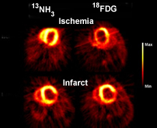
- The above images compare13NH3 and FDG in the ability to identify ischemia/infarct. Based on the available data13NH3 seems to be the superior agent for cardiac viability. Question - Would13NH3 be considered an appropriate radiopharmaceutical for a cardiac stress/rest procedure
- 15Oxygen-water =
- Properties

- T1/2 = 2 minutes
- Used to analyze brain and heart
- In-house cyclotron required
- H215O and/or C15O are used
- Biodistribuiton
- General
- Diffuses easily into tissue making it an excellent perfusion agent
- Used to assess cerebral and myocardial blood flow (MBF). Gold standard for assessing blood flow
- Repeated images can be done over a short period of time to evaluate different oxidative states (ex. different types of brain stimulation)
- Cardiac
- In general, this agent continues to increase in its extraction fraction as blood flow increase and can reach ~100%
- Stress and rest images cannot be done in one imaging session because its T1/2 is to short
- 15O water can be administered, however, high levels of activity remain in the vascular pool
- If radiolabeled water is administered blood volume images must be corrected with C15O. For this reason other PET cardiac agents are recommended
- Brain
- Used to analyze blood flow to the brain
- n-15O-butanol
- Can be used to analyze blood flow to the brain as well as flow to other areas of the body
- Consider the partition coefficient of 15O - water is 0.9 where 15O-butanol is 1.0
- What does partition mean? It is compound's solubility, hence, butanol has better solubility and may be "slightly" better agent in evaluate MBF
- 82Sr-82Rb generator =
- Properties
- Parent - 82Sr has a 25.6 day T1 /2
- Daughter - 82RbCl has a 76 second T1/2
- 0.02 :Ci/mCi breakthrough limit which is checked daily, prior to patient use
- Is administered through an infusion system
- Extraction fraction = 50-60% (however, difficult to assess correctly)
- Biodistribution
- Evaluates blood flow to the myocardium
- Is an analog to potassium and is picked up by myocardial tissue via active transport
- Uptake increases with increased blood flow increases, however, at high flow rates the extraction levels off
(just like thallium)
- For the above reason, quantification is not recommended
- It has poor blood clearance in the blood pool and has a limited T1 /2 . Therefore, activity is present within the cardiac chambers even as it perfuses into cardiac tissue. This requires a subtraction technique when images are processed
- According to Stankewicz, MA, et. al. 82Rb is not a good predictor for myocardial viability
- Other ways to image the Brain
- Evaluation of neurotransmission
- Evaluation of the dopaminergic nervous system with respect to neurotransmitter and neuroreceptor
- Nerves send messages by the production of neurotransmitters that diffuse across a synaptic cleft which then binds to a receptors on a postsynaptic nerve
- This neurotransmitter pathway works in two ways
- Inhibits transmission - stops the pulse
- Excites a transmission - starts it or is allows the pulse to continue
- Reduced function of the neurotransmitters translates to certain types of disease (examples - Huntington's and Parkinson's disease)
- PET can be used to to evaluate dopaminergic pathways
- In addition, the effectiveness of antipsychotic drugs can be assessed with PET
- Several PET agents have been used to evaluate nerve conduction and the D2 receptor sites
- 18F-fluorodopa (FD)
evaluates enzymatic activity =
- Dopamine is a neurotransmitter
- Assess the number of dopaminergic neurons in the brain
- Levodopa is a pharmaceutical that converts to dopamine and is given to patients to elevate dopamine levels within the brain. Patients suffering from low levels of dopamine usually have some form of neurological disorder (ex. Huntington)
- FD follows the same pathway as levodopa and interact with aromatic amino acid decarboxylase to form (enzymatic process)
- FD (flourodopa)
 fluorodopamine
- Levodopa Dopamine
- Fluorodopamine then accumulates around the neurons containing D2 receptors
- Degenerative diseases show reduced uptake of the radiopharmaceutical around the basal ganglia and based on the intensity of its uptake severity of disease can be determined
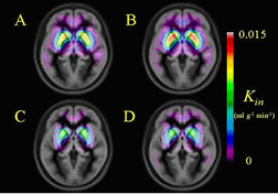
- The above image demonstrates the level of FD uptake that is co-registered with an MR scan: (A) Considered normal brain activity, (B) After treatment with levodopa, (C) Parkinson's disease after medication has been stopped, and (D) medication of levodopa has resumed
- 11C-RAC (rapclopride) evaluates neuroreceptors
- RAC has a high affinity and high specific activity with D2 receptor in the brain
- In response to low levels of dopamine up-regulation occurs at the synapse sites were there is an increase in the amount of receptors. The opposite occurs with down-regulation where dopamine levels are high and the amount of receptor sites are reduced
- How does this effect the uptake of RAC?
- First, consider the concept of competitive inhibition, where the RAC is competing with D2 receptor sites
- In Parkinson's disease upregulation occurring, therefore there is an increase in D2receptor sites and a decrease in dopamine production. Therefore, RAC uptake would be considerable, where FD would be reduced
- In psychotic disorders usually there is down-regulation with a high level of neurotransmitters being released
http://brain.oxfordjournals.org/cgi/reprint/120/12/2187
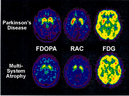
- From the above images present Parkinson's Disease and Multi-system atrophy. Note the difference between the diseased and the type of radiopharmaceutical being acquired. When evaluating these images consider: dopamine, D2 receptors, and glucose uptake
- In general what would happen to 11C-RAC or 18F-FD with up vs, down-regulation and in the increase vs. decrease levels of neurotransmitter dopamine?
- Which radiopharmaceuticala are used to image what?
- Cardiac
- Perfusion
- 82Rb
- K analog - Na - K pump
- blood flow
- 13N-H3
- Alkaline medium
- Glutaminic pathway
- Brain and heart
- 15O Water
- Blood flow to brain, heart, or tumor
- Flow and oxygen utilization
- Viability
- 18FDG
- Fake sugar
- phosphorylates with hexakinase and then trapped because it cannot dephosphorylate (sugar can)
- At 1 hour post dose tumor activity increases over inflammation
- Suggest 90 delay for best results for target to background and SUV calculation
- 11C plamitate
- 13N-glutamate
- Fatty acid metabolism - 11C-palmitate
- Brain
- Metabolism: 18FDG
- Oxygen utilization: 15O2
- Receptor: 18F - 6-fluoro-L-DOPA
- Many others
- Oncology
- Glucose: 18FDG
- Synthesis of amino acids:11C-acetate
- Hypoxia: 18F-fluoromisonidazole (FMISO)
- Binds to intracellular macromolecules that are in a hypoxic state
- Cell proliferation: 18F-fluorothymidine (FLT)
- Cell proliferation
- Perfusion: 15O water
- Lipids: 11C-choline
- Bone - Na18F
Return to the Beginning of the Document
Return to the Table of Content
For a more complete list of radiopharmaceuticals developed for PET visit the Athinoula A. Martinos Center for Biomedical Imaging
-
References - Radiopharmaceuticals in Nuclear Pharmacy and Nuclear Medicine by Kowalsky RJ and Falen SW, 2nd edition, 2004.
- Molecular Anatomical Imaging. von Schulthess, GK, 3rd edition 2003
- Quantification of fatty acid uptake with [11C]palmitate-PET, 2007-11-29 Vesa Oikonen- Draft
- Kinetic models for analyzing myocardium [11C data. Hugo, WA, et al. Eur J Nucl Med (2009)
- Utility of 18F-FDG PET in Evaluating Cancers of Lung, Acker, MR, et al, JNMT (2005)
- Clinical Applications of PET in Brain Tumor, Geffen, D, et al. JNM (2007)
updated - 11/30/09