- In theory (and technically) it depends on the arrival of the two 511 keV photons to each specific detector
or element
- Usually the time window is set to between 4 to 12 nsec
- When the two gammas arrive within the window the event is recorded
- Distance can then determined by defining which one arrives first, which one arrives second
- By having this data you should be able to extrapolate back the origin/location of the annihilation event
- In actuality TOF is being applied with current PET technology, however, it has its limitation. Let us discuss the TOF process
and its limitations
- Given the right software/hardware the timing window must be set to 0.6 nsec. Question - how does this effect the system's sensitivity?
- At tissue with this technology is the inability to place the exact point of the annihilation event, however, we can get close and it's within 9cm
- Initially one might think that 9cm is just too much to make a real imaging difference. But this type of thinking will get you into trouble!
- While spatial resolution of a PET systems is in the range of 5 to 10 mm, the fact that we can pinpoint the event, via TOF, to 9cm means that we can more accurate place the LOR within a very limited area
- Without TOF all LORs must be back-projected through their entire path
- With TOF all LORs can be backprojected to length that is no greater than 9cm
- Furthermore, because a patient's transaxial slice is significantly greater than 9cm
- The end result is that smaller LORs improve spatial resolution
- Let us also think about the size of the body habitus, application of TOF, and scatter
- The larger the patient and the lack of TOF results in increased scatter. The fact remains, the larger the diameter these gammas travel through the greater probability of Compton, further increasing the scatter events
- If TOF is applied this improves image quality via the reduction of scatter. In fact, the larger the patient the more effective TOF will be, as this relates knowing, within 9cm, where the annihilation event occurred
- How does scatter and size relate? Without the application of TOF a 120 kg patient's S/N ratio would be 6 times greater when compared to someone who weighed 50 kg. In addition the larger patient, because of attenuation, requires an increase in scanning time to acquire acceptable count density. It is suggested that when TOF is applied to the larger patient image quality improves and the concern of the 6 to 1, S/N ratio, no longer applies
- What components does a PET Scanner require to apply the TOF?
- Crystals must have a high light output
- LSO and LYSO (yttrium impurity) accomplish this. When compared to BGO, BGO does not have a high light output
- Because of the improved light output the time window can be reduced from 4 nsec to 0.6 nsec
- PMTs and related electronics in today's technology are faster allowing to process of data in shorter time intervals
- Image acquisition and reconstruction is done in 3D list-mode with iterative reconstruction being applied. This allows for better spatial and temporal resolution, however, the computer crunching is very intensive
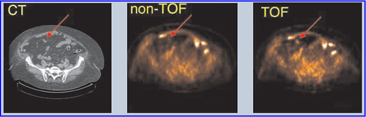
- The above data shows the application of TOF in a 3-D PET image. Note the red arrow is pointing to an additional "hot spot" not seen in the "non-TOF." TOF seems to definitely improve image quality, based on the images above.
Information for this outline was attained from SNM Center of Excellence Newsletter/Fall 2006, Time-of-Flight PET by Joel S. Karp, PhD
Return to the Table of Content
Next Lecture - Introduction to CT Instrumentation