Case Studies with Bone Imaging
Cancers of the Bone
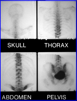
- Case 1 - Fluid uptake in the related to pleural effusion caused by a malignant hepatoma. In addition the patient also has cirrhosis. This image was initially copied from Dartmouht-Hitchcock.org who's link is no longer active
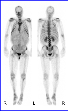
- Case 2 - As an example of photopenia this patient was believed to have Chordoma, a malignant tumor that arise form remnant embryonic cells of the vertebra. It usually attacks the spine and coccyx. http://www.med.harvard.edu/JPNM/BoneTF/Case19/WriteUp19.html
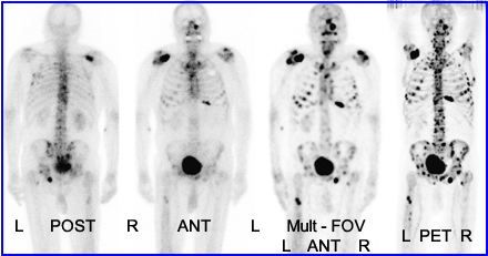
- Case 3 - When comparing MDP to Na18F with patients that have metastatic prostate cancer it appears that PET is more sensitive in finding metastatic tumor (Even-Sapir, et al). This research article evaluates of WB Bone with MDP, WB Bone multi-FOV SPECT, and PET Na18F. PET found more lesions and has improved sensitivity and specificity when compared to MDP 70/57% and 92/82% respectively. http://jnm.snmjournals.org/cgi/content/full/47/2/287/FIG1
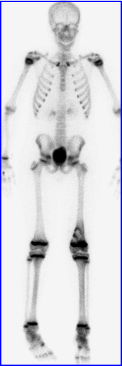
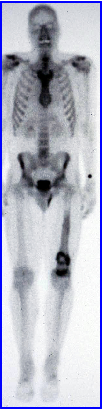
- Case 4 - Osteosarcoma is usually seen in younger patients and occur in about 400 cases per year in the US. http://www.med.harvard.edu/JPNM/CH/JS3/WriteUp.html
- Case 5 - Chondrosarcoma is noted in the distal portion of the femur. This type of cancer has a 600 per year incidence in the US. Usually found where there is cartilage. http://sarcomahelp.org/chondrosarcoma.html
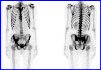
- Case 6 - Twenty year old male was diagnosed with Ewing's Sarcoma. Statistically its is found in lower limb (45%), pelvis (25%), Upper limb (13%), spine/rib (13%) http://radiopaedia.org/cases/ewing-sarcoma-pelvis
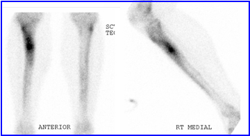
- Case 7 - Osteoid Osteoma - This 22 year old male had complained of shin pain for a three month period which was thought to be associated with his running activities. An apparent stress fracture was seen on x-ray. After testing with CT, MRI, and Nuclear Medicine, it was biopsy disease was confirmed.
Return to Table of Content
The Detection of Bone Metastases in Patients with High-Risk Prostate Cancer: 99mTc-MDP Planar Bone Scintigraphy, Single- and Multi-Field-of-View SPECT, 18F-Fluoride PET, and 18F-Fluoride PET/CT. by Even-Sapir, Metser, Mishani, et al.. 2006 JNM