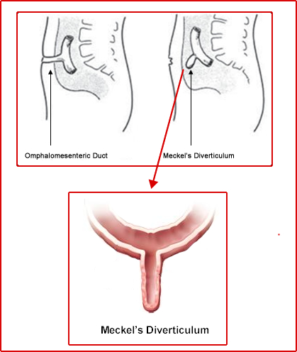
Meckels Diverticulum
- Anatomy and Physiology
- Is a congenital abnormality that occurs in about 2% of the pediatric population
- Is caused is a bulge in the lower portion of the small intestine and a congenital defect. Essentially it is a left over form the umbilical courd
- Usually found in the antimesenteric side of the small bowel and approximately 46 to 67 cm from the ileocecal valve
- Ranges between 1 to 12 cm in size
- Approximately 25% of the time it contains gastric mucosal cells
- Pain and GI bleeding is usually associated with this abnormality
- Patient is usually asymptomatic
- Bleeding is caused by HCl and pepsin secreted from ectopic gastric mucosa causing ulceration to the adjacent tissue
- Rules of 2s
- Occurs in 2% of the population
- Is usually within 2 feet of the ileocecal valve
- Average length is 2 inches
- Patient is usually symptomatic by the age of 2
- Pathophysiology of the radiopharmaceutical
- 99mTcO4- is used
- Pertechnetate has an affinity for mucosal cells
- If Meckel's is present, when pertechnetate is injected a hot spot in the lower abdomen is seen
- Procedure
- Patient should be NPO 12M
- Patient is placed in the supine position
- Camera/Computer setup
- 256 x 256 matrix
- Collimator LEHR or LEGap
- Five minute acquisitions
- Place the camera head in the anterior projection over the abdomen
- Inject 200 μCi/kg of 99mTcO4- IV
- Take 1 image every 5 to 10 minutes for an hour
- Display data
- Pharmacologic intervention can further enhance detection
- Glucagon promotes pooling and prevents pertechnetate from moving down the GI track (dose 50 to 100 μg)
- Pentagastrin increases pertechnetate uptake (dose 6 μg/kg)
- Cimetidine as a histamine antagonist and blocks secretion of pertechnetate from the gastric mucosal cells (dose 300 mg QID for 1 2 days)
- Ranitidine just like cimetidine, however, there are fewer side effects (dose 1 mg/kg given IV over 20 minutes: start the procedure 1 hour post dose)
- Case Review
- Case 1
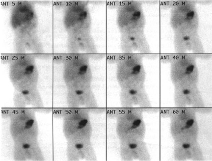
- The above example is of a normal Meckels scan taken over 60 minutes
- Note the distribution of pertechnetate
- Stomach becomes very intense with activity why?
- Significant background is seen throughout the body why?
- Notice how the myocardium initially appears very intense, but decreases in activity over time why?
- Case 2
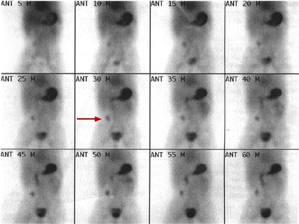
- The above is an example of an abnormal study
- Notice that over time an increase of activity is seen in the lower right quadrant of the abdomen (red arrow)
- This is the location of the Meckels Diverticulum
- Sensitivity/Specificity
- Sensitivity is 85% or greater
- Specificity is 95%
- Main reason for a false negative exam is the lack of gastric muscosal cells in the diverticulum
Meckels Diverticulum Procedure
Return to the beginning of the document
Return to the Table of Contents
11/21