Cavernous Hemangioma
- This type of liver disease was mentioned previously in your liver/spleen lecture and now requires a more in-depth look
- Anatomy and physiology
- Found in the liver usually in the right lobe
- Lesion contain large blood volume, endothelial-lined spaces, and may be more diffused than be capsulated
- Occurs in 3 to 7% of the population and has a greater affinity in women (4:1)
- Usually benign and singular, however, can appear multiple
- Is usually located in the right lobe of the liver
- Usually the patient is symptomatic, however, if the tumor gets too large it can displace the surround structure and the patient may have interment pain
- Usually found accidentally in a CT scan nuclear medicine is used to determine the nature of the lesion
- Procedure
- Inject 20 to 30 mCi of technetium labeled RBCs
- Dynamic and immediate static images will show an area within the liver void of uptake
- Two to three hour delayed images will show increased activity within the cold area seen in the initial images
- SPECT imaging is suggested
- LEHR collimator
- 360 degree
- 128 x 128 matrix
- 120 stops at 30 seconds per stop
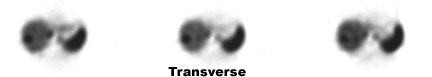
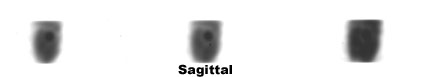
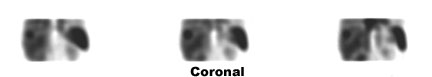
- Case Review
The following SPECT images show an area of increased uptake in the right lobe of the liver. This is seen in all three views: transverse, sagittal, and coronal. If a Tc99mSulfur Colloid study was done in the same venue a lack of uptake would be noted in the same projections.
Return to the Table of Content