Red Cell Survival - Procedure
- With A-C-D solution from the A-C-D tagging vial, wet a 20 mL syringe and withdraw 15 mL of blood from the patient.
- Slowly and gently (to prevent hemolysis) aseptically inject the contents of the syringe into the vial of A-C-D solution.
- Add 7.4 MBq (200 µCi) of Sodium Chromate :51Cr into the A-C-D mixture.
- Gently mix the blood by intermittent swirling every 5 to 10 minutes.
- Allow to tag at room temperature for 30 minutes.
- Withdraw 20 mL of the labeled RBC from the A-C-D vial and re-inject it IV into the patient.
- Twenty-four hours post injection withdraw 10 mL of patient blood. This becomes the "day zero" of the procedure and the considered the standard
- Every 2 to 3 days later remove a 10 mL sample and continue for at least 28 - 30 days
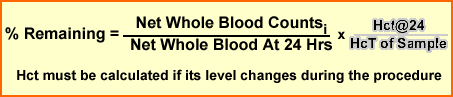
- Determine the hematocrit of each sample and if there is a variation then this factor should be applied to the calculation below
- Issues regarding each sample
- Each sample should be labeled with the date and time of withdrawal.
- Each withdrawal should be at approximately the same time each day.
- Frequency of sampling depends primarily on convenience.
- For statistical accuracy a minimum of 10 samples should be obtained.
- Pipette 4 mL of each sample into a counting vial and label accordingly.
- Count all samples at the same time to negate the effect of radioactive decay.
- The calculations are based on using the 24-hour sample as 100% and making it the starting point. All other samples are calculated as a percent of the 24-hour sample, and indicate the percent remaining. If later samples have hematocrits different from the 24-hour sample, the correction below should be made.
- After calculating each individual sample the percent remaining should be plotted on the logarithmic scale with the Y-axis being % Remaining and X-axis being time (in days). Draw a best-fit straight line through the points.
- The red cell survival time is determined from the graph by finding the time at which the straight line reached 50 percent.
- Normal Range: Normal 28 to 40 days
- Abnormal Less than 28 days
Comments
- The labeling process for this procedure is very similar to RCV, a couple of exceptions
- Don't add ascorbic acid to the ACD vial
- Plasma and Whole body standards do not have to be made - the patient's 24 sample becomes the standard
- If you stop the procedure before 28 days and apply regression analysis to the curve to determine the T1/2 survival, your data may be inaccurate. I speak from an actual case were regression define the T1/2 to be greater than 28 days, but the physician decided to continue with the procedure and when we counted the 28th day sample less than 50% of the RBCs had survived.
- If the patient requires blood transfusions then
- Make sure that it is done just before you start the RCS procedure
- Do not let the patient have a transfusion until the RCS procedure is completed - otherwise it will lower your survival curve.
Return to the Table of Content
The U.S. Nuclear Regulatory Commission has approved distribution of this radiopharmaceutical to persons licensed to use byproduct material listed in Section
35.100, and to persons who hold an equivalent license issued by an Agreement State.
1 Kocher, David C., “Radioactive Decay Tables,” DOE/TIC-11026, page 74 (1981).
2 Method of Calculation: “S” Absorbed Dose per Unit Cumulated Activity for Selected Radionuclides and Organs, MIRD Pamphlet No. 11 (1975).
Revised/ 6/97
Mallinckrodt, Inc.
St. Louis, MO 63134