Procedure on Dual Isotope Imaging Using Indium and MDP
Imaging infection using 99mTcMDP and 111InWBC
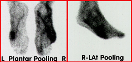
- Following the injection 25 mCi of 99mTcMDP pooling images are taken of the feet (see above). The patient has a suspected infection following amputation of several toes
- 500 uCi of 111InWBC was then injected into the patient and delayed images were taken the following day

- Images are collected using both the 99mTc peak and 111In peak (see above)
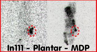
- ROIs are then drawn around the area of interest (see above)
- Maximum pixel count in the 99mTc ROI is 1219
- Maximum pixel count in the 111In ROI is 602
- A ratio is identified (1219/602) as 2.02 which is used for normalization of the indium
- Applying image normalization and summing
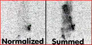
- In order to normalize the indium counts all indium pixel counts are multiplied by 2.02 (see above)
- 99mTcMDP images and normalized 111InWBC images are summed (see above)
- Note that there is additional area of focal uptake identified when the images are fused
Return to the beginning of the document
Return to the Home Page