Hematopoietic System - Bone Marrow

  1. There are three types of tissue found in this system

      Where do Bone Marrow Cells Come From?

      http://en.wikipedia.org/wiki/Hematopoietic

    1. Myeloid
      1. Part of the hematopoietic system that contains bone marrow, which produces: red blood cells, white blood cells, and platelets
      2. Production occurs specifically by myeloid tissue
    2. Lymphoid
      1. Found in several parts of the body, which includes: mucous membrane of the GI track, respiratory track, lymph nodes, spleen, and thymus
      2. This is where differentiation of lymphocytes occur (ex. plasma and memory cells - more on that later in the semester)
      3. However, the stem cells that produce lymphoid's and myeloid's are found in the bone marrow
    3. Reticuloendothelial system (RES)
      1. These cells are not produced by Hemocytoblasts, do not play a role in production of cells for circulation, however, RES and myeloid tissues share the same space within the bone marrow
      2. "Cleaning the blood" through a process known as phagocytosis

    4. The Portal Triad - Remember this when studying liver spleen imaging?
  • Growth and development of the bone marrow
    1. Every bones in a new born child contain active red marrow and RES, located in the central portion of bone, defined as medullary canals
    2. Between the ages of 2-5 yellow marrow begins develop within the distal portions of the skeletal system (hands, feet, and distal tibia) and progress inwardly
    3. Between the ages of 5-10 further regression of the red marrow is seen. Usually 1/2 of the tibia contains yellow marrow, while active marrow is still noted throughout the femur
    4. After the age of 10 and throughout a person's adult life active red marrow will be located in the central skeleton and the proximal ends of the humerus and femur
    5. Hence, based on age, yellow marrow replaces red bone marrow through a child's development and this process is completed around the age of 10
    6. Therefore the level of erythropoiesis (red cell production) relates to the amount of red bone marrow. The great the amount of red marrow, the more RBC, white, and platelets production occurs
    7. Applying the right radiopharmaceutical, will assist in determining the amount of red marrow uptake, defining possible disease related to bone marrow
  • A look at the different radiopharmaceuticals used to image the hematopoietic system
    1. Ideally iron would be the number one choice for bone marrow evaluation since iron is required in the production o RBCs
      1. 52Fe is a positron emitter that has an 8.2 hour half-life. The half-life essentially allows us to ship it in time to almost any location within the US, however, availability is limited and the cost is expensive. In addition, a PET scanner is required for imaging. Oh and what about reimbursement through insurance
      2. 59Fe has two energy gammas one at 1.1 MeV and the other at 1.3 meV with a 45 day half-life. Can we use this radiotracer?
      3. 111In-chloride is considered an analog to iron and adhere to plasma transferrin (iron behaves in a similar manner) and therefore may be a suitable replacement for iron. However there are limitations to consider if In is used
        1. Once indium enters the vascular pool it tags to transferrin and has a biological T1/2 of about 6 hours, yet iron biological T1/2 is only 1-2 hours.
        2. 111In uptake in the liver and spleen is significantly greater than radio-iron. When liver/spleen uptake is extreme activity will interfere the ability to resolve data in that region
        3. Animal studies indicate that iron has greater uptake in red marrow when compared to indium
        4. In comparison with 59/52Fe, 111In was:
          1. 59Fe was more sensitive in identifying bone marrow production in patients' that received radiation treatments2
          2. 111In showed several false positives with Polycythemia Vera and anemia (compared with 59Fe2
          3. When there is a dissociation between colloid and RBC production, 111In was found to be a poor agent to image the hematopoietic system3
        5. Finally, indium's uptake properties are more similar to radio-colloid and than with radio-iron
      4. Since bone marrow and RES share the same sinusoids within bone 99mTc-sulfur colloid can be considered
    2. Of the four radiopharmaceuticals used sulfur colloid seems to be the "best" approach
  • Imaging procedures
  • Normal Bone Marrow Scan with Tc99mSC

    1. 99mTc-sulfur colloid procedure - The above image1 shows normal biodistribution of 99mTcSC within the RES. Bone marrow is noted in the central skeleton with no peripheral expansion. In regards to image quality liver/spleen is not excessively "hot" allowing for excellent analysis of bone marrow distribution in and around this area
      1. Procedure
        1. Collimator - LEPH
        2. 20% window
        3. 10 - 15 mCi given IV (concept - overload the REC system with radio-colloid)
        4. Wait at least 15 minutes before imaging (what is you see cardiac uptake?)
        5. Use a slow scan speed (6 - 10 cm/minute) scan and image head to toe
      2. In1111 scan with Nomral (L) and Abnormal (R)

    2. 111In-chloride4
      1. Collimation - ME
      2. Photopeaks
        1. 171 keV @ 20%
        2. 245 keV @ 20%
      3. Delayed whole body imagine can be done at 24 to 48 hours
      4. Scan speed should be no greater than 6 cm/minute
      5. Scan head to toe
    3. 59/52Fe procedures
      1. 59Fe cannot be accurately imaged with today's technology. Why? At best one might evaluate via an uptake probe, but even that would give limited data
      2. 52Fe is a positron emitter which is only evaluated in research facilities
    4. Caution!! While RES and red marrow (erythropoietic) share the same space, distribution of colloid and iron can differ!5 Several patients showed different uptake between the three radiopharmaceuticals. Those patients had Hodgkin, Polycythemia Vera, and Myelofibrosis (refer to the chart on page 366)
    5. Another interesting link related to bone marrow uptake with Sulfur Colloid - https://link.springer.com/chapter/10.1007/978-3-319-56167-7_8
  • Diseases
    1. Aplastic anemia
      1. Disease shows failure within the bone marrow that produces red/white cells and platelets
      2. Usually there is an increase in yellow bone marrow that may invade the red bone marrow within the central skeleton
      3. While disease may be the cause (viral hepatitis), certain drugs or chemicals (benzene, choramphenicol, quinine, etc.) it has also been related to the lack of iron in the diet
      4. Lose of bone marrow uptake would be an indication of Aplastic anemia
    2. Pattern with Thrombocytopenia, myelocytic leukemia, and Polycythemia Vera
      1. Initially the central skeleton looks normal
      2. As disease progresses, there is failure within the central skeleton to produce the appropriate cell components that causes expansion of the red marrow into the axial skeleton and peripheral long bones
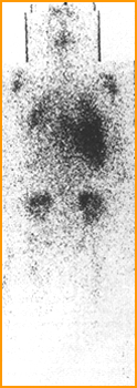
      3. Further progression of the disease will show increased splenic uptake when 52Fe is administered, however, there is no increase in spleen size
      4. Chronic Lymphocytic Leukemia Polycythemia Vera with Bone Marrow Expansion
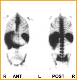
      5. The left image1 shows activity extending down into the the patient's knees as well as extensive spleen uptake. This is an example of chronic lymphocytic leukemia and the radiopharmaceutical used was 52Fe
      6. The image1 on the right is a classic example of Polycythemia Vera taken with 52Fe. In this case the activity extends down to the knees and shows reduced uptake in the central skeleton
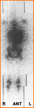
    3. Sickle cell anemia

    4. https://www.mayoclinic.org/diseases-conditions/sickle-cell-anemia/symptoms-causes/syc-20355876

      1. Genetically altered red blood cells can cause infarct and ischemia with bone tissue/marrow as well as other areas of the body
      2. In general, infarct/ischemia is not seen in the bone marrow, however, increased activity is noted in the axial skeleton and peripheral bones
      3. Two Types of Sickle Cell Anemia

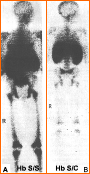
      4. Two types of sickle cell anemia1 are identified above
        1. Hb F is fetal hemoglobin that becomes Hb A (adult) and occurs in about 95% of the population
        2. Hb S/S is a form of sickle cell hemoglobin that shows extensive homogeneous activity in the peripheral and axial skeleton. In addition the liver and spleen may be enlarged
        3. Hb S/C is another variation of sickle cell that shows similar involvement (axial and peripheral bone) but with reduced uptake
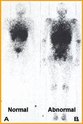
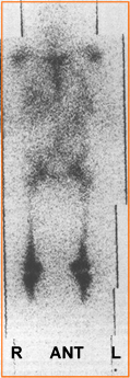
    5. Whole Body Radiation Therapy
      1. This causes loss of bone marrow production, but over time bone marrow function returns. Usually activity in the hemopoietic system starts up within a several months after therapy and normal distribution should return within the year
      2. Bone Response to Whole Body Radiation3

      3. The above image1 is an example of a patient that received a total of 4000 rads. At least 6 months post therapy 52Fe shows the return of red marrow production with a marked increased to the splenic area. The literature was not clear as to why there was significant spleen activity

    Return to the beginning of the Document
    Return to the Table of Content

    1 - Images acquired from Diagnostic Nuclear Medicine, 3rd Edition, MP Sandler, et. al., vol 2, Williams and Wilkins, Baltimore, 1996

    2 - Comparative study of 111In and 59Fe bone marrow scanning by Parmentier, C, et al., European Journal of Nuclear Medicine

    3 - A comparison of 111In with 52Fe and 99mTc-sulfur colloid for bone marrow imaging by Merrick, MV, et al., JNM

    4 - Bone-Marrow Imaging with Indium-111 Chloride in Aplastic Anemia and Myelofibrosis: Concise Communication by Sayle, BA, et al. JNMT

    5 - >Differences in Distribution of Erythropoietic and Reticulendothelial Marrow in Hematologic Disease by Dyke DV, et al Blood

    9/23