CEA-Scan Finds Disease Missed by
Both CT & Pet in an Asymptomatic
Patient with Rising Serum CEA
PATIENT HISTORY:
A 44 year-old female with right colon cancer resected in May 1998 followed by a hysterectomy in December of 1998. Chemotherapy started in August of 1998 continues at the time of this evaluation.
SERUM CEA LEVELS: 38 ng/ml
PRIOR DIAGNOSTIC STUDIES:
Negative abdominal/pelvic CT (May 1999)
Negative abdominal/pelvic PET (June 1999)
SPECT Imaging (June 1999) identifies focal activity in the right pelvis superior to the bladder.
SURGICAL FINDINGS:
Metastatic disease in the right pelvis.
CONCLUSION:
CEA-Scan can change patient management by identifying the source of rising serum CEA levels missed by both CT and PET.
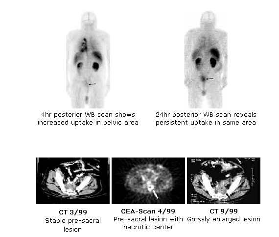
CEA-Scan resolves ambiguous CT, negative colonoscopic biopsy
RJ is a 57-year-old male with a history of infiltrating adenocarcinoma of the colon confirmed by colonoscopic biopsy and resected 11 days later.The patient did well for almost 3 years, when he had rectal bleeding. Colonoscopy revealed what appeared to be friable tumor but histology was negative. An abdominal x-ray performed a week later was negative.
The patient underwent a CEA-Scan study three days later. The scan was positive for a tumor on the left side of the pelvis (circled in transaxial image at right, coronal view at left).
A chest CT was negative. A CT of the abdomen and pelvis (below) performed the same day revealed nonspecific hypodensities in the liver and mild mural thickening (circled area) within the sigmoid colon which was reported as "may be secondary to post-surgical changes/underfilling with contrast, but cannot definitely exclude recurrent neoplasm."
An exploratory laparotomy and low anterior resection the following week found no palpable liver lesions and a 4-cm adenocarcinoma at the rectosigmoid junction. Thus, CEA-Scan correctly identified the source of the bleeding in an area that was equivocal on both CT scan and colonoscopy.
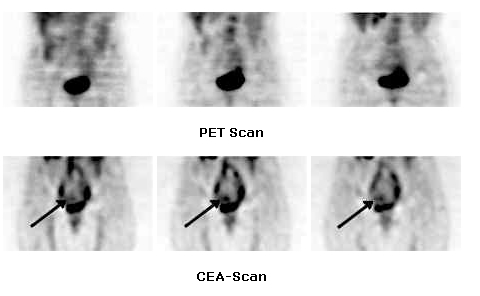
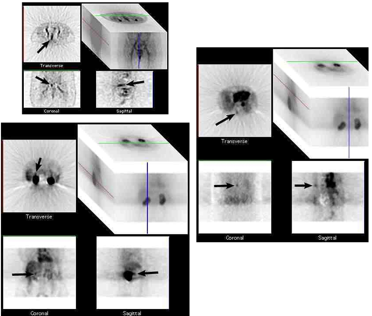
CEA-Scan Helps Find Distant Metastasis in a Patient with Primary Colorectal Cancer and a Suspected Artifact on CT
These CEA-Scan images are of a 72-year-old female complaining of constipation and tiredness accompanied by anemia. Colonoscopy showed a lesion 20-cm from the anal verge and left-sided diverticulosis. The CT scan showed the lesions seen with the colonoscopy, an aortocaval node and what appeared to be a volume-averaging artifact in the right upper quadrant that may have been caused by respiration. SPECT images with CEA-Scan confirmed all the lesions found with the CT and colonoscopy, including the primary tumor (image block 1). CEA-Scan also showed uptake in the same right upper quadrant area as the suspected artifact on CT (image block 2) clearly demonstrating that the density seen on CT was indeed a lesion and not an artifact. Both findings were surgically confirmed. This case is an example of how CEA-Scan can provide additional information and helps find metastasis often not easily appreciated with other modalities.
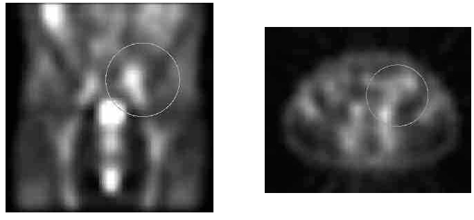
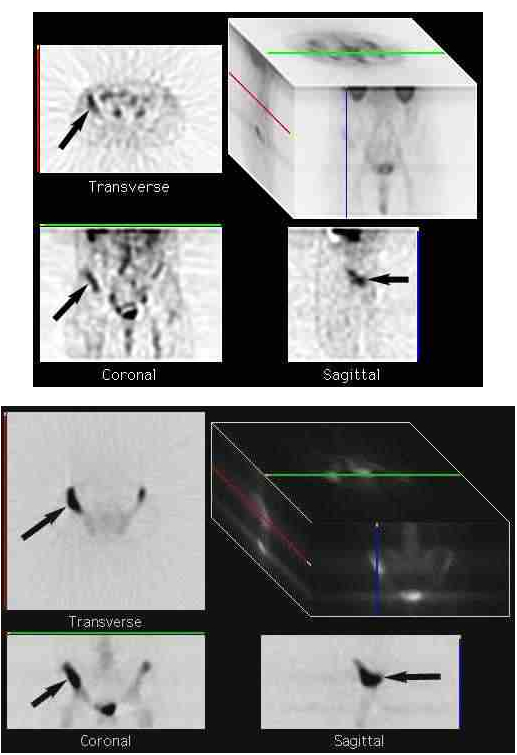
CEA-Scan Helps Identify Osseous Metastasis in the Pelvis of a Patient with a History of Colorectal Cancer and Rising Blood CEA levels
These CEA-Scan images are of a 66-year-old male with a history of right sided colon cancer. He has had no signs or symptoms of recurrence for over 2 years but now presents with a rising blood CEA. SPECT images with CEA-Scan at 3 hours post injection revealed uptake along the right iliac crest (image block 1), which was also present in the 24 hour images (not shown). In order to determine if the suspicious pelvic uptake is in the soft tissues adjacent to the ileum or in bone, a radionuclide bone scan was performed 6 days later. The SPECT bone scan showed intense accumulation of the radiopharmaceutical in the right iliac wing (image block 2) correlating the findings on the CEA-Scan. This suspected abnormality had not been detected with other anatomical imaging studies.
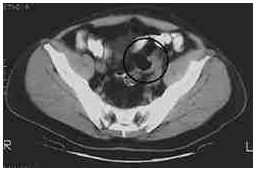
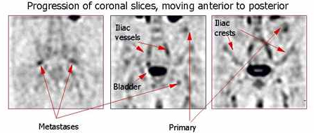
CEA-Scan Helps Stage Patient with Primary Colorectal Cancer
This is a series of coronal CEA-Scan images of a 58-year-old male who presented at the ER of his hospital with clear evidence of obstruction. The resident on duty ordered a CT study, which clearly showed the probable site of obstruction as a primary colon tumor, and areas of hypodensity in the liver, suggestive of metastatic disease. However, no lymph node involvement was noted on the CT. The CEA-Scan showed extensive lymphatic involvement, and metastatic disease in the liver. The slide calls out several landmarks: The bladder and kidneys are normally intense, because they're the organs of excretion of the antibody fragment and isotope. The iliac vessels, iliac crests and genitalia are visible due to normal blood pool. This case illustrates how CEA-Scan provides information unduplicated by CT in the extrahepatic abdomen and pelvis, and helps confirm suspicious regions in the liver.
Data (text and images) was copied from http://www.cea-scan.com/cases/scan.htm