Journal of Comprehensible Results
Sanchez E, Bigbee J, Fobbs W, Robinson S, Sato-Bigbee C (2008)
Opioid Addiction and Pregnancy:Perinatal Exposure to Buprenorphine
Affects Myelination in the Developing Brain.
Glia 56:1010-1027
Opioid Addiction and Pregnancy:Perinatal Exposure to Buprenorphine
Affects Myelination in the Developing Brain.
Glia 56:1010-1027
(Translated by Brenna Kent)
Experiment: Number, Diameter, and G Ratio of Axons
|
|
|
|
Buprenorphine not only has the potential to affect the composition and timing of myelination, but also the caliber of the axons and the ratio of myelinated to unmyelinated axons.
In order to determine how axons were affected, they were examined in samples from 26 day old rats which represents the end of the myelination period. The number of myelinated axons, the diameter of axons, and the G Ratio were found. The G Ratio is calculated by dividing the diameter of the naked axon by the diameter of the myelinated axon, so a larger G Ratio indicates a smaller myelin sheath(Fig. 8). |
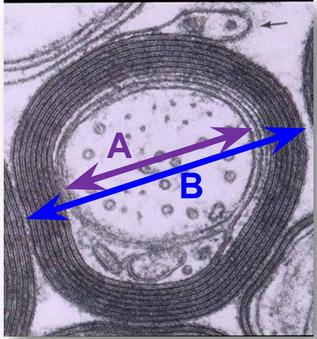
Figure 8: Representation of how the G Ratio is calculated: A: Diameter of naked axon B: Diameter of myelinated axon G Ratio= A/D |
|
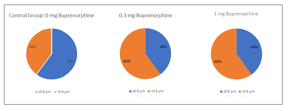
There was a significant decrease in the number of myelinated axons in rats who were exposed to 1 mg/kg/day of buprenorphine in utero (Fig 9). It was also found the exposure to buprenorphine only affected the diameter of myelinated axons and that unmyelinated axons had a diameter similar to that of the control. For both treatment groups, myelinated axons had a larger diameter (Fig 10). Not only did the diameter of the myelinated axons increase, but there was also a decrease in the relative thickness of the myelin sheath.
The fact that only the diameter of myelinated axons and not unmyelinated axons were affected could indicate that this difference is being caused by the affect of buprenorphine on the oligodendrocytes since they would have interacted with myelinated axons. Altering the diameter and thickness of the myelin sheath could impact the speed at which electrical impulses could travel through axons. |
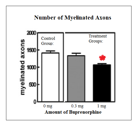
Fig. 9: Examination of the number of myelinated axons. In the 1 mg/kg/day of buprenorphine group there was a significant decrease in the number of myelinated axons which is indicated by the red star. Adapted from "Opioid Addiction and Pregnancy:Perinatal Exposure to Buprenorphine Affects Myelination in the Developing Brain." by Sanchez E, Bigbee J, Fobbs W, Robinson S, Sato-Bigbee C, 2008, GLIA, 56, p. 1021. Copyright 2008 by Wiley-Liss, Inc.
Adapted from "Opioid Addiction and Pregnancy:Perinatal Exposure to Buprenorphine Affects Myelination in the Developing Brain." by Sanchez E, Bigbee J, Fobbs W, Robinson S, Sato-Bigbee C, 2008, GLIA, 56, p. 1022. Copyright 2008 by Wiley-Liss, Inc. |