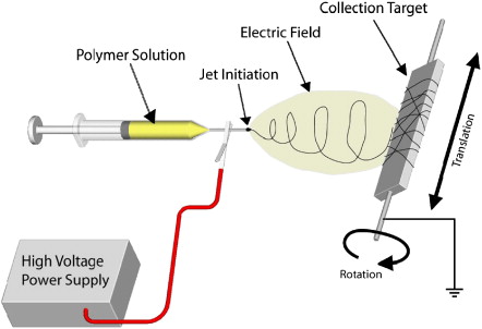
Electrospinning
Electrospinning traces its roots to electrostatic spraying, which was first described more than 100 years ago. In electrostatic spraying, charge is injection into a liquid, typically 5-30 kV, from an electrode. The charged liquid is separated some distance from a second electrode (target) of opposite polarity to establish a static electric field. A so-called Taylor Cone forms due to the competing forces of the static electric field and the liquid’s surface tension. For liquids with a finite conductivity, charged droplets are dispersed from the tip of the Taylor Cone and are delivered to the target. If the liquid consists of a polymer melt or a polymer in solution and the concentration of that polymer is sufficiently high to cause molecular chain entanglement, a fiber, rather than a droplet, is drawn from the tip of the Taylor cone.
A basic electrospinning system consists of a charged polymer solution (or melt) that is fed through a small opening or nozzle (usually a needle or pipette tip). Because of its charge, the solution is drawn toward a grounded collecting plate (usually a metal screen, plate, or rotating mandrel), typically 5 – 30 cm away, as a jet. During the jet's travel, the solvent gradually evaporates, and a charged polymer fiber is left to accumulate on the grounded target. The charge on the fibers eventually dissipates into the surrounding environment. The resulting product is a non-woven fiber mat that is composed of tiny fibers with diameters between 50 nanometers and 10 microns. This non-woven mat forms the foundation of the scaffold. If the target is allowed to move with respect to the nozzle position, specific fiber orientations (parallel alignment or a random) can be achieved. Previous work has shown that the mechanical properties of the scaffold can be varied by varying the fiber diameter and orientation.
Basic Electrospinning set up
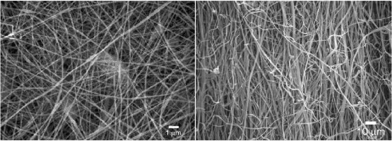
Some electrospinning examples
-
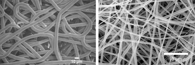
Collagen
Fibril forming collagens, type I, II and III are the most abundant proteins found in the body. The collagen nanostructure provides appropriate biological signals/space to the cells for growth, development and repair. In addition, collagen provides the overall structural integrity and strength to tissues. Our laboratory has fabricated nanofibrous collagen scaffolds that retain biological and structural properties. The electrospun collagen fiber diameters ranged from 100 nm to 5 microns.
Type I collagen with randomly oriented fibers (left) and aligned fibers (right).
-
Elastin
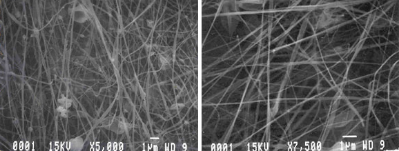
Elastin is a highly insoluble, hydrophobic protein that is required by the body in organs where shape and energy recovery are crucial. It is found largely in the walls of arteries and veins to provide elastic recoil and to prevent dissipation of transmitted pulsatile energy into heat. Our lab has successfully electrospun solubilized elastin at concentrations ranging from 200 mg/ml to 333 mg/ml, which resulted in the creation of flat ribbon like fibers.
Scanning electron micrograph of electrospun elastin at 250 mg/ml.
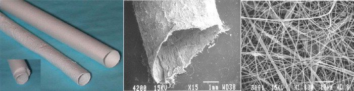
Photograph of an electrospun composite structure (Left), Scanning electron micrograph of tubular electrospun composite (Middle), Scanning electron micrograph of electrospun blend of collagen type I, collagen type III, and elastin (Right) .
-
Fibrinogen
Fibrinogen is a soluble protein found in blood plasma that participates in wound healing as nature's provisional matrix.
Fibrous structure of electropsun human fibrinogen is illustrated in the pictures above.
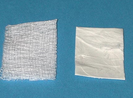
Photograph of cotton gauze (left) and bovine electrospun fibrinogen mat (right) for potential use as a tissue engineering scaffold or wound dressing.
-
Polydioxanone
PDO is a highly crystalline, biodegradable polyester that was originally developed for use as a degradable suture (Ethicon, Inc., a Johnson and Johnson Company). PDO's excellent biocompatibility, strength, elasticity, flexibility and shape memory are desirable for the creation of future engineered vascular grafts.
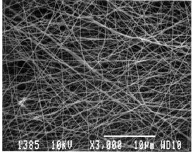
Scanning electron micrograph of 180 nm diameter randomly oriented fibrous structures produced from 42 mg/ml PDO (above).
-
Poly(lactic acid)
PLA is a biodegradable aliphatic polyester. Our lab uses PLA synthesized from the more common L optical isomer (often denoted PLLA) instead of the D or dl isomers. This is because the product of PLA hydrolysis (l-lactic acid) occurs naturally in the metabolic pathway in humans.
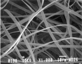
Scanning electron micrograph of randomly oriented PLA electrospun from chloroform (Left) and HFP (Right).
Most content on this page is from the article in Advanced Drug Delivery Reviews, 59(14), 1413-1433, 2007.