BLOOD, A SPECIALIZED CONNECTIVE TISSUE

BLOOD AND HEMOPOIESIS

Readings: Basic Histology, 10th Ed., by Junqueira, and Carnerio. Chapt. 12 and 13. Color Atlas of Basic Histology, Berman; Blood (8) and Bone marrow (9). Web page: Best results with Netscape >4.78.

OBJECTIVES: To teach you to identify blood cell types, and some cells of the hematopoietic series.

I. GENERAL CHARACTERISTICS OF BLOOD

The blood shares many of its characteristics with connective tissues (CT), therefore, it is often classified as a specialized, atypical CT; e.g.:

A. Common characteristics of CT vs. Blood

CT:   Blood:  
1. Various types of cells:
1. Cells:
Erythrocytes (RBCs)
      Leukocytes (WBCs)
      (Particles: Platelets
      Chylomicrons=1m diam. Fat droplets)
       
2. Few cells per unit volume:
2. Cells:
45%; + Plasma: 55%;
       
3. Intercellular matrix:
3. Blood plasma:
  a. Fibers:   a. Fibrin: forms fibers upon clotting only;
  b. Ground substance:   b.Serum: the fluid separated from clotted blood;

B. Quantitative characteristics of blood (Expressed as mean figures)

1. 7% of body weight;
2. 5 quarts (~5 liters) per 150 pound man;
3. Erythrocytes: 5,000,000 in females and 5,500,000 in males per 1 cubic millimeter of blood;
4.
Leukocytes: Range: 5,000 to 9,000 per cu. mm of blood in adults; Below and above these limits disease is indicated.
5. Ratio of WBCs to RBCs is 1:700
6. Blood platelets: Range: 200,000 to 400,000 per cubic millimeter.


C. CLASSIFICATION OF BLOOD CELL TYPES

Based on color in vivo and after staining in vitro.

(NOTE: "Classification" -- means: The grouping of blood cells according to their morphological and staining differences. So, a neutrophil would be classified as a "granular leukocyte" so would the eosinophil or the basophil. Lymphocytes and monocytes would be classified as "agranular leukocytes." Erythrocyes are classified as "erythrocytes," so identification and classification for RBC's is the same. If you are asked to identify a granulocyte, then you would have to write its name e.g., basophil, eosinophil or neutrophil. Similarly with the agranular leukocytes, e.g., a lymphocyte may be identified as a small or medium lymphocyte.)
II. MORPHOLOGY, STAINING AND FUNCTIONS OF BLOOD ELEMENTS

A. Erythrocytes

1. Size: 7.5 µm in diameter

2. Morphology:

a. Nucleus: No nucleus;

b. Cytoplasm: Biconcave disk; 95% of its mass is hemoglobin (Hb); Hb imparts a red color in vivo;

3. Function: Transport O2 and CO2; (Total RBC surface area for gas exchange is 42,000 sq. yds.)

4. Physical properties: a. soft, flexible, elastic;

b. Rouleaux -- surface tension;

c. Crenation -- hypertonicity;

d. Hemolysis -- hypotonicity;

e. Agglutination -- agglutinins (antibodies);

OOOOOOOOOOOOOOOOOOOOOOOOOOOOOOOOOOOOOOOOOOOOOOOOOOOOOOOOOOOOOOOOOOOOOOOOO SLIDE 33. Go to the virtual slide box ( http://www.path.uiowa.edu/virtualslidebox/ ) . Search for blood, normal, and look at the three slides numbered 33. Blood smear (Wright's Stain). The majority of cells seen on this slide are Erythrocytes. These cells are stained light red. Observe that their center is lighter stained. This is due to the near absence of the cytoplasm in the center where the biconcavity is located. On some slides some of the RBCs look like sweetgum balls, spiny (see diagram on previous page), these RBC are crenated (shrunken and spiny) due to loss of water (you may not see this on the virtual slides-- check). OOOOOOOOOOOOOOOOOOOOOOOOOOOOOOOOOOOOOOOOOOOOOOOOOOOOOOOOOOOOOOOOOOOOOOOOO
B. Leukocytes - possess ameboid motility

1. Granular Leukocytes: have abundant specific granules in their cytoplasm. Named: "specific granules" because their staining is unique for the cell.

a. Basophils: 0.5% of WBCs

Size: 9 - 12µm diameter;

Nucleus: U or S shaped;

Cytoplasm: Round, spherical in vitro; Filled with coarse, basophilic, membrane bound, water soluble granules which contain: (over)

(1). Heparin
(2). Histamine
(3). Slow-Reacting substance of Anaphylaxis (SRA-A);
(4). Eosinophil Chemotactic Factor A (ECF-A);

Function: involved in allergic and inflammatory reactions.

OOOOOOOOOOOOOOOOOOOOOOOOOOOOOOOOOOOOOOOOOOOOOOOOOOOOOOOOOOOOOOOOOOOOOOO For basophils go to the Digital Histology CD. Blood smear (Wright's Stain). Basophils are larger than the surrounding RBCs (see above). Difficult to find. Why? The cytoplasm contains many distinct dark purple, metachromatic granules. (What is metachromasia? - look it up!). Nucleus is S or U shaped, lighter purple, usually partially masked by the granules. Observe and be able to recognize the ultrastructure of basophils (see basic histology text).
OOOOOOOOOOOOOOOOOOOOOOOOOOOOOOOOOOOOOOOOOOOOOOOOOOOOOOOOOOOOOOOOOOOOOOO

b. Eosinophils: 3% + of WBCs

Size: 9 - 12 µm diameter;

Nucleus: bilobed in human, annular in rodents, relatively heterochromatic.

Cytoplasm: Round, spherical in vivo;

Contains coarse, eosinophilic (acidophilic), membrane bound granules; Contain a crystalloid (seen with EM only). The granules are primary lysosomes containing acid hydrolyzes.

Function: Participate in allergic reactions by phagocytizing Ag-Ab complexes and by this dampen the allergic response. Do not ingest bacteria.

OOOOOOOOOOOOOOOOOOOOOOOOOOOOOOOOOOOOOOOOOOOOOOOOOOOOOOOOOOOOOOOOOOOOOOOO SLIDE 30. Blood smear (Wright's Stain). Eosinophils stain red (dirty red sometimes). The eosinophilia is due to the numerous coarse granules which fill the cytoplasm. The nucleus consists of two round to oval lobes connected by a nearly invisible bridge of the nucleus. This nuclear morphology is diagnostic of eosinophils. If tissue is obtained from rodents the nucleus is doughnut shaped (annular). Be able to identify the diagnostic crystalline ultrastructure of eosinophil granules as shown in electrom micrographs.
OOOOOOOOOOOOOOOOOOOOOOOOOOOOOOOOOOOOOOOOOOOOOOOOOOOOOOOOOOOOOOOOOOOOOOOO

c. Neutrophils: 65% + of WBCs

Size: 10 - 12µm diameter;

Nucleus: Lobated, 3-5 interconnected lobes; polymorphonuclear ("polys"); Clumped chromatin.

Cytoplasm: Round, spherical in vivo;

Contains two types of granules:

a. Numerous small specific granules with low staining affinity; Contain phagocytins (bactericidal).

b. Few, large azurophilic granules (lysosomes); these contain: hydrolytic enzymes and myeloperoxidase.

Function: Phagocytize bacteria and particulate matter in CT, then die and form pus.


OOOOOOOOOOOOOOOOOOOOOOOOOOOOOOOOOOOOOOOOOOOOOOOOOOOOOOOOOOOOOOOOOOOOO Virtual SLIDE 33. Search for normal Blood (neutrophils). Blood smear (Wright's Stain). Neutrophils have 3-5 dark purple staining nuclear lobes. Cytoplasm contains very small reddish granules and sometimes some even smaller azurophilic granules. The background staining of the cytoplasm tends to be pale pink. Most numerous of WBCs.
OOOOOOOOOOOOOOOOOOOOOOOOOOOOOOOOOOOOOOOOOOOOOOOOOOOOOOOOOOOOOOOOOOOOO


2. Agranular Leukocytes: their granules are few and small; seen only by EM.

a. Lymphocytes: 25% of WBC

Size: 7-8µm diameter;

Nucleus: Round, heterochromatic, may or may not show a small indentation.

Cytoplasm: Round, spherical in vivo, scanty, appears as a thin, lightly basophilic rim around nucleus of small lymphocytes; Medium lymphocytes have more; Large lymphocytes are rarely found in blood; Occasionally have few azurophilic granules (lysosomes);
Function: In immune responses; T and B lymphocytes; B lymphocytes can differentiate into Ab-producing plasma cells in CT and lymphoid tissues.

OOOOOOOOOOOOOOOOOOOOOOOOOOOOOOOOOOOOOOOOOOOOOOOOOOOOOOOOOOOOOOOOOOOOO Virtual slide 33. Search for normal blood. Blood smear (Wright's Stain). Lymphocytes in blood smears: 1. Small L.: Dark purple heterochromatic nucleus surrounded by a very thin rim of pale blue cytoplasm (usually seen only on one side). 2. Medium L.: Larger, have visible cytoplasm all around. 3. Large lymphocytes: are the same in appearance but about twice as large as small lymphocytes - usually not seen in blood smears. Always compare cell size to the erythrocyte for reference. Lymphocytes in sections: even a small lymphocyte has cytoplasm all around the nucleus. OOOOOOOOOOOOOOOOOOOOOOOOOOOOOOOOOOOOOOOOOOOOOOOOOOOOOOOOOOOOOOOOOOOOO


b. Monocytes: 5% ± of WBCs

Size: 9-17µm diameter;

Nucleus: Large with a deep indentation; Kidney or U shaped, heterochromatic;

Cytoplasm: Round to oval, abundant; Contains: scattered azurophilic granules (Lysosomes);

Function: After differentiating into a macrophage in the CT, it becomes phagocytic;

OOOOOOOOOOOOOOOOOOOOOOOOOOOOOOOOOOOOOOOOOOOOOOOOOOOOOOOOOOOOOO Virtual SLIDE 33. Search for normal blood. Blood smear (Wright's Stain). The monocyte is the largest cell in a blood smear. Have a medium blue chromatin (kidney or U shaped nucleus), gray blue cytoplasm and sometimes fine, lilac-colored (azurophilic) granules. OOOOOOOOOOOOOOOOOOOOOOOOOOOOOOOOOOOOOOOOOOOOOOOOOOOOOOOOOOOOOO

C. Platelets: are cytoplasmic fragments of megakaryocytes of the spleen and bone marrow.

Size: 2-3 µm in length;

Shape: Round to oval, flattened, from side view rod shaped;

Nucleus: none;

Cytoplasm: Contains deeply basophilic, centrally located granules. This region is called the granulomere; Granules are surrounded by a pale staining cytoplasmic rim, the hyalomere.

Function: Adhere to injured regions of vessels and help in clotting of the blood. May release clotting factors to enhance fibrin formation. Can release fibrinolysin to dissolve clots. Contain serotonin and other vasomotor agents causing vascular contraction. Below normal levels (200,000/cu. mm.) the endothelium of blood vessels becomes leaky and hemorrhagic.

OOOOOOOOOOOOOOOOOOOOOOOOOOOOOOOOOOOOOOOOOOOOOOOOOOOOOOOOOOOOOOO Virtual SLIDE 33. Search for normal blood and platelets. Blood smear (Wright's Stain). Platelets appear indistinguishable at low magnification from debris on blood smears. At high power (X100, oil), platelets have violet to purple granulomere and light blue hyalomere. Tend to be found in groups. Can you recognize the ultrastructure of platelets?
OOOOOOOOOOOOOOOOOOOOOOOOOOOOOOOOOOOOOOOOOOOOOOOOOOOOOOOOOOOOOO

D. Megakaryocyte (Note: normally located in the bone marrow and spleen red pulp)

Size: 50 -70 µm in diameter;

Nucleus: Multi-lobed; have a coarse chromatin pattern;

Cytoplasm: Contains fine azurophilic granules; like in platelets;

Function: Gives rise to platelets by cytoplasmic fragmentation;

OOOOOOOOOOOOOOOOOOOOOOOOOOOOOOOOOOOOOOOOOOOOOOOOOOOOOOOOOOOOOOOOO
Virtual SLIDE 34: Search for normal bone marrow smear and click on the radio buttons. (Human bone marrow smear; Wright's Stain). Be able to identify a megakaryocyte in bone marrow mears (lone slide) and in decalcified, H&E-stained sections (Slide # 23). In both smears and sections of bone marrow look for the largest cells at 10x. These will be megakaryocytes. In slide #23 megakaryocytes are immature and typically have only two lobes. Compare these cells in sections with those in the bone marrow smear. What are two distinguishing characteristics of megakaryocytes? (Size and multilobed nucleus).
OOOOOOOOOOOOOOOOOOOOOOOOOOOOOOOOOOOOOOOOOOOOOOOOOOOOOOOOOOOOOOOOO

Practice the characteristics of the formed elements of the blood by filling out the chart on this page (use the figures for numbers and sizes of cells given in this syllabus)

Cells & Particles
Abundance
Size
Shape of Nucleus
Cytoplasmic content

Function

RBCs          
Eosinophils          
Basophils          
Neutrophils          
Lymphocytes          
Monocytes          
Platelets          


III. HEMOPOIESIS: BLOOD CELL FORMATION

A. Phases

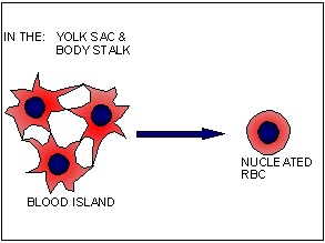
1. Mesoblastic - (begins at 3rd week of gestation)

a. embryonic;

b. in yolk sac and body stalk;

c. condensation of mesenchy-mal cells - form blood islands;

d. nucleated blood cells form

2. Hepatic - (begins 4-8 weeks of
gestation)

a. in liver, thymus, and spleen;

b. in lymph nodes somewhat latter;

c. forms anucleated RBCs

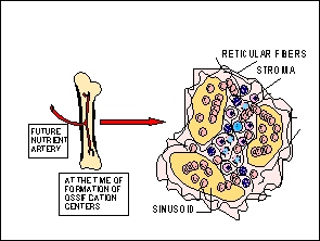
3. Myeloid - (starts at 12 weeks of gestation)

a. in bone marrow;

b. major site of hemopoiesis;

c. begins with establishment of ossification centers in bones;

d. all blood cell types found in adults can be produced by the bone marrow;

B. Myeloid Tissue

1. Formation

2. Stroma

a. a meshwork of reticular fibers;

b. perivascular cells are multipotent;

c. vascular supply;

(1) nutrient arteries

(2) sinusoids

d. differentiation of blood cells takes place in the stroma;

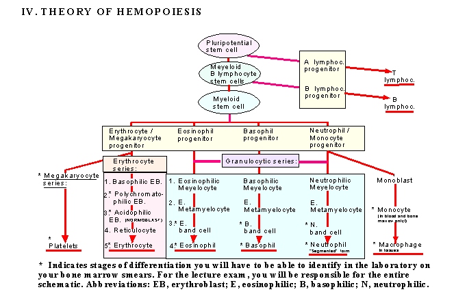
3. Production / Differentiation of blood elements - Terminology;

(1) Erythropoiesis - Erythrocytes

(2) Granulopoiesis - Granulocytes

(3) Lymphopoiesis - Lymphocytes

(4) Monopoiesis - Monocytes

(5) Thrombopoiesis - platelet formation from megakryocyte;

OOOOOOOOOOOOOOOOOOOOOOOOOOOOOOOOOOOOOOOOOOOOOOOOOOOOOOOOOOOOOOOOOOOO
Human Bone Marrow smear (Wright's Stain). Study the bone marrow on virtual slide 34 (click on the radio buttons) and study the cells from the CD provided. Bone marrow smears contain all stages of cell differentiation found in the bone marrow. Note that there are a lot more nucleated cells on bone marrow smears than on the blood smears. Blood smears contain only mature cells. You need to learn to identify those stages of cell differentiation which are indicated by an asterisk (*) on the chart above.

Identify: (See CD for reference)

A. In the erythrocytic series:

1. Polychromatophilic erythroblast - Cell size: nearly twice the size of an RBC. Nucleus: Round, dark purple with condensed heterochromatin; Cytoplasm: Irregular, sometimes ragged outline; Cytoplasm is stained bluish gray with spots of lightly acidophilic (pinkish) areas which show the beginning of hemoglobin synthesis.

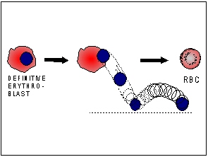
2. Acidophilic erythroblast (Normoblast) - Cell size: only slightly larger than a mature RBC. Nucleus: small, acentric, dark purple and highly condensed (pyknotic). Cytoplasm: Round and stains acidophilic (light red), almost the same color as the staining of the RBCs. Sometimes the nucleus is seen being ejected from the cytoplasm.

3. Erythrocytes - as before.

B. In the granulocytic series:

Identify the band forms (e.g., Eosinophilic, Basophilic, and Neutrophilic band cells) of the different granular leukocytes. These are characterized by the presence of either elongated (sausage-shaped) or horseshoe-shaped nuclei. The cytoplasm contains the specific granules of the particular series. So if the granules are red then the cell is called the eosinophilic band cell etc. You should also find the mature or segmented nuclear forms of neutrophils, the bilobed nuclear form of eosinophils, and mature "polys."

C. Among the agranular leukocytes:

1. Monocytes - as in the blood smear;
2. Macrophages - will be identified in CT latter.

Virtual SLIDES 10, AND 22. Search for normal bone. Decalcified, H&E-stained, bone sections with bone marrow. Identify the bone marrow and study its composition. Classify the epithelia lining the sinusoids.
Identify normoblasts and megakaryocytes (largest cell in the bone marrow; can be identified at low magnification). What are two distinguishing characteristics of megakaryocytes?

OOOOOOOOOOOOOOOOOOOOOOOOOOOOOOOOOOOOOOOOOOOOOOOOOOOOOOOOOOOOOOOOOOOOOOO