Electrospinning traces its
roots to electrostatic spraying, which was first described more than 100 years
ago. In electrostatic spraying, charge
is injection into a liquid, typically 5-30 kV, from an electrode. The charged liquid is separated some distance
from a second electrode (target) of opposite polarity to establish a static electric
field. A so-called Taylor Cone forms
due to the competing forces of the static electric field and the liquid’s
surface tension. For liquids with a
finite conductivity, charged droplets are dispersed from the tip of the Taylor Cone and
are delivered to the target. If the
liquid consists of a polymer melt or a polymer in solution and the
concentration of that polymer is sufficiently high to cause molecular chain
entanglement, a fiber, rather than a droplet, is drawn from the tip of the Taylor cone.
A basic electrospinning system
(Figure below on left) consists of a charged polymer solution (or melt) that is
fed through a small opening or nozzle (usually a needle or pipette tip). Because of its charge, the solution is drawn
toward a grounded collecting plate (usually a metal screen, plate, or rotating
mandrel), typically 5 – 30 cm
away, as a jet. During the jet's travel,
the solvent gradually evaporates, and a charged polymer fiber is left to
accumulate on the grounded target. The
charge on the fibers eventually dissipates into the surrounding
environment. The resulting product is a
non-woven fiber mat that is composed of tiny fibers with diameters between 50
nanometers and 10 microns. This non-woven
mat forms the foundation of the scaffold.
If the target is allowed to move with respect to the nozzle position,
specific fiber orientations (parallel alignment or a random) can be
achieved. Previous work has shown that
the mechanical properties of the scaffold can be varied by varying the fiber
diameter and orientation.
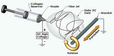
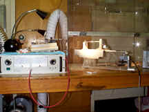
Schematic representation of an electrospinning device
(left) and photograph of the
current system used in our laboratory.
Collagen Electrospinning Images
Elastin Electrospinning Images
Fibrinogen Electrospinning Images
Synthetics Electrospinning Images
Cellular
interaction with Electrospun Collagen-Elastin
http://www.people.vcu.edu/~glbowlin/electrospinning.htm
2006-8-19